
Fig. 2
- ID
- ZDB-IMAGE-080401-36
- Genes
- Publication
- Ignatius et al., 2008 - colgate/hdac1 repression of foxd3 expression is required to permit mitfa-dependent melanogenesis
- All Figures
- Figures for Ignatius et al., 2008
|
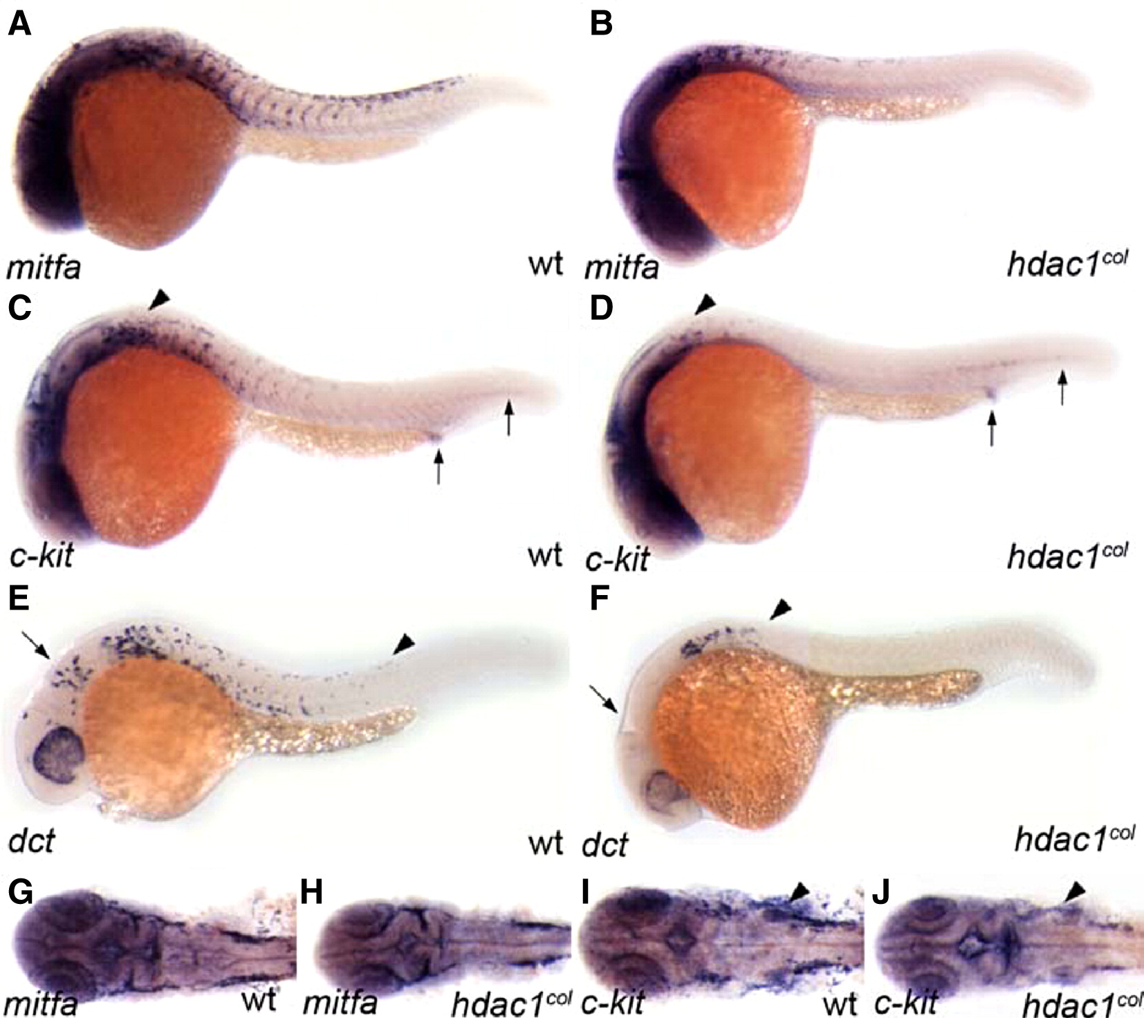
Fig. 2 Fewer melanoblasts are specified in hdac1col embryos. Lateral and dorsal (cranial region) views of 25 hpf embryos that are stained by in situ hybridization to reveal expression of mifta (A, B, G, H), c-kit (C, D, I, J) and dct (E, F). (A, B, G, H) There are fewer mitfa-positive melanoblasts specified in hdac1col mutants as compared to wild type. (C, D) Melanoblast-specific c-kit expression in hdac1col mutants is absent and/or reduced (arrowheads), although non-melanoblast expression of c-kit in the post anal region and posterior mesoderm is equivalent to wild-type (arrows). (I, J) Similarly, in the cranial region melanoblast-specific c-kit expression is reduced or absent, while c-kit expression in the branchial arches is more robust albeit disorganized in hdac1col mutants as compared to wild-type (arrowheads). (E, F) There are fewer dct-positive differentiating melanoblasts in hdac1col mutants compared to wild-type (arrowheads) and most of the dct-positive melanoblasts are located posterior to the otic vesicle in the dorsal stripe suggesting defects in migration. Also, there is a lack of dct-positive melanoblasts in the anterior head (arrow).
Reprinted from Developmental Biology, 313(2), Ignatius, M.S., Moose, H.E., El-Hodiri, H.M., and Henion, P.D., colgate/hdac1 repression of foxd3 expression is required to permit mitfa-dependent melanogenesis, 568-583, Copyright (2008) with permission from Elsevier. Full text @ Dev. Biol.