
|
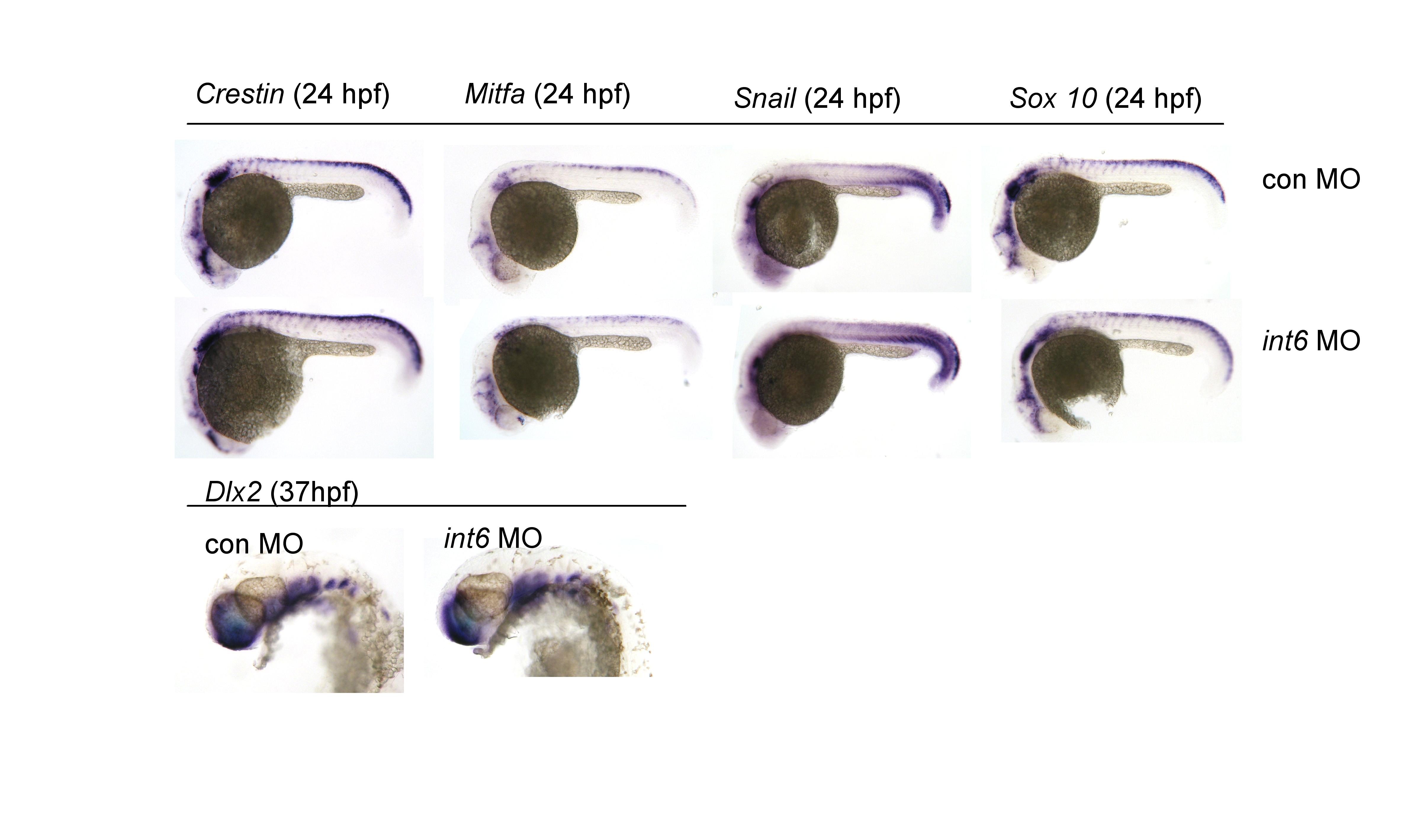
Fig. S2 Lateral views of in situ hybridization of neural crest markers in control and int6 morphants, revealing no change in cell number or migration as indicated by the apparently normal expression of dlx2 (stages 6–36 hpf, examined at two hour intervals), nor of early markers of NCC and melanocytes, such as sox10, crestin, snail and mitfa (24 hpf) in int6 morphants. These observations were extended by examination of a transgenic sox10-GFP line (1) revealing unaltered GFP-expressing NC-derived cells in int6 morphants within the first 48 hpf, but a loss of GFP expressing differentiated pharyngeal arches 3–7 by 3 dpf (data not shown).