
Image
|
Figure Caption
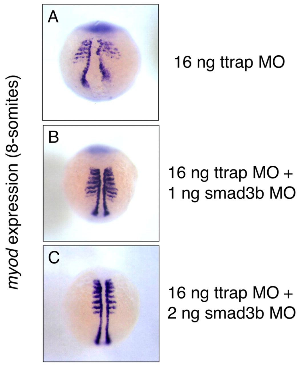
Fig. 8 Double Ttrap and Smad3b knockdown rescues CE defect in Ttrap knockdown. myod marks paraxial/adaxial mesoderm (8 ss). (A) Broadened somites and a wide distance between myod cells in Ttrap single knockdown embryo. (B) A combination of 16 ng TtrapMO and 1 ng Smad3bMO shows closer convergence of myod cells at midline. However, somitic expression is still broad relative to the fully rescued embryo co-injected with 2 ng Smad3bMO (C), which now displays the normal myod domain. Dorsal views, anterior at top.
Figure Data
Acknowledgments
This image is the copyrighted work of the attributed author or publisher, and
ZFIN has permission only to display this image to its users.
Additional permissions should be obtained from the applicable author or publisher of the image.
Full text @ Development