
|
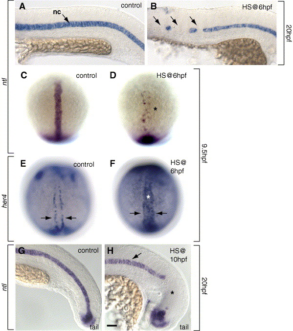
Fig. 4 Notch signaling limits notochord and promotes hypochord development during gastrulation. (A, B, G, H) Side views, anterior to the left, at 20 hpf (22-somite stage) and (C, D) dorsal views, animal pole at top, at 9.5 hpf (YPC stage) showing ntl-expressing notochord cells. (A) Control embryo showing normal pattern of ntl-expressing notochord at the midline. (B) Expression of NICD at 6 hpf reduced trunk notochord (arrows). (C) Normal pattern of notochord precursors at 9.5 hpf. (D) The number of notochord precursors was reduced (asterisk) in embryos after induction of NICD at 6 hpf. (E, F) her4 expression in hypochord precursors at 9.5 hpf, dorsal view. (E) her4 was expressed in a row on both sides of the midline (arrows) in control embryos. (F) her4 was expressed throughout the midline (asterisk) after expression of NICD at 6 hpf. (G) Control embryo showing normal pattern of tail ntl expression in the notochord. (H) Expression of NICD at 10 hpf reduced tail notochord (asterisk), but trunk notochord appeared normal (arrow). Scale bar: 40 μm for panels A, B, G, H; 80 μm for panels E, F.
Reprinted from Developmental Biology, 298(2), Latimer, A.J., and Appel, B., Notch signaling regulates midline cell specification and proliferation in zebrafish, 392-402, Copyright (2006) with permission from Elsevier. Full text @ Dev. Biol.