
|
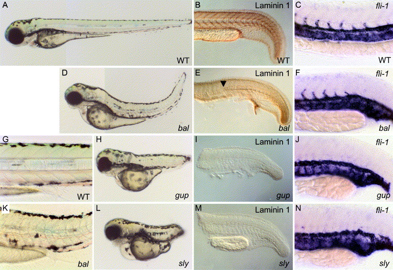
Fig. 2 Morphological and molecular analysis of bal, gup and sly mutant embryos. Lateral views show wild-type (WT) (A, G), bal (D, K), gup (H) and sly (L) embryos at 3 day post fertilisation. Staining with a polyclonal antibody generated against mouse laminin 1 (α1β1γ1) detects expression pattern in 24 hpf WT (B) embryos. Laminin immunoreactivity in 24 hpf bal mutant embryos (E) is at wild-type levels within the posterior notochord (arrowhead), but severely reduced immunoreactivity is seen in the non-differentiated anterior notochord, and intersomitic boundaries. In 24 hpf gup (I) and sly (M) embryos, laminin 1 immunoreactivity is abolished. The developing vasculature is marked by expression of fli-1 mRNA in 24 hpf WT (C) and bal (F), gup (J) and sly (N) mutant embryos.
Reprinted from Developmental Biology, 289(1), Pollard, S.M., Parsons, M.J., Kamei, M., Kettleborough, R.N., Thomas, K.A., Pham, V.N., Bae, M.K., Scott, A., Weinstein, B.M., and Stemple, D.L., Essential and overlapping roles for laminin alpha chains in notochord and blood vessel formation, 64-76, Copyright (2006) with permission from Elsevier. Full text @ Dev. Biol.