
Image
|
Figure Caption
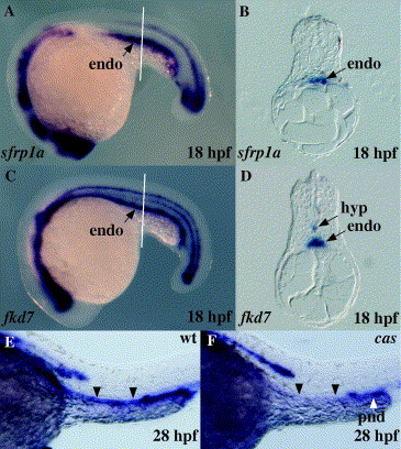
Fig. 3 Analysis of sfrp1a expression in the developing gut. Whole mount in situ hybridisation was performed with an antisense RNA probe for sfrp1a (A, B, E and F) or fkd7 (C and D) at the indicated stages. Embryos are presented in whole mount with anterior to the left (A, C, E and F), or in 5 μm transverse sections (B and D) at the level indicated in A and C (white lines), respectively. A wild-type embryo is compared to a cas mutant embryo (E and F). The wild-type location of endodermal tissue is indicated with arrowheads. endo, endoderm; hyp, hypochord; pnd, pronephric duct.
Figure Data
Acknowledgments
This image is the copyrighted work of the attributed author or publisher, and
ZFIN has permission only to display this image to its users.
Additional permissions should be obtained from the applicable author or publisher of the image.
Reprinted from Gene expression patterns : GEP, 6(8), Pezeron, G., Anselme, I., Laplante, M., Ellingsen, S., Becker, T.S., Rosa, F.M., Charnay, P., Schneider-Maunoury, S., Mourrain, P., and Ghislain, J., Duplicate sfrp1 genes in zebrafish: sfrp1a is dynamically expressed in the developing central nervous system, gut and lateral line, 835-842, Copyright (2006) with permission from Elsevier. Full text @ Gene Expr. Patterns