
|
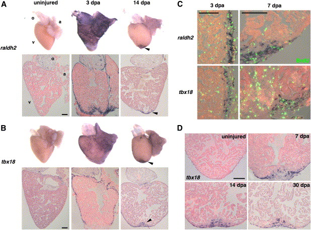
Fig. 3 Organ-Wide Activation of Epicardial Cells and Invasion of the Regenerate (A and B) Whole-mount (top) and section (bottom) ISH of uninjured and 3 and 14 dpa hearts for raldh2 (A) and tbx18 (B). Expression of each marker is low or undetectable in the uninjured hearts but induced at 3 dpa in epicardial tissue surrounding the atrium (a) and ventricle (v) for tbx18 and these tissues plus the outflow tract (o) for raldh2. The endocardium surrounding wounded myofibers also expresses raldh2. By 14 dpa, expression of both markers is localized to the apical wound (arrowhead). (C) Colocalization of BrdU (green) with raldh2 (top) and tbx18 (bottom) in the ventricular epicardium at 3 and 7 dpa. The left images display a lateral edge of the 3 dpa ventricle away from the wound, while the right images display a lateral and apical portion of the 7 dpa ventricle including some of the wound. By 7 dpa, raldh2/tbx18/BrdU-positive cells begin to localize to the injury site. The majority of nonepicardial BrdU-positive cells are erythrocytes within the ventricular lumen. (D) Epicardial-derived cells positive for tbx18 invade the regenerate by 7 dpa and remain by 30 dpa. Scale bar = 100 μm.
Reprinted from Cell, 127(3), Lepilina, A., Coon, A.N., Kikuchi, K., Holdway, J.E., Roberts, R.W., Burns, C.G., and Poss, K.D., A Dynamic Epicardial Injury Response Supports Progenitor Cell Activity during Zebrafish Heart Regeneration, 607-619, Copyright (2006) with permission from Elsevier. Full text @ Cell