
IMAGE
Fig. 4
Image
|
Figure Caption
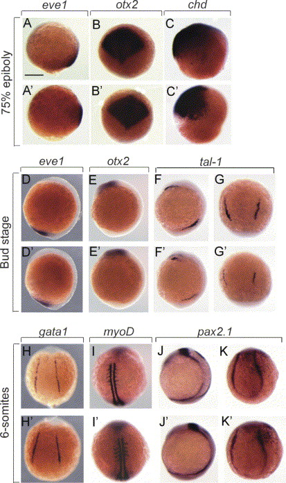
Fig. 4 Analysis of dorsoventral markers on crim1 morphants. (A–K′). Wholemount RNA in situ hybridisation of 8–12 hpf embryos injected with 5 ng control-MO (A–K) and crim1A-MO (A′–K′) and stained with eve1 (A, A′, D, D′), otx2 (B, B′, E, E′), chordin (C, C′), tal1 (F, F′, G, G′), gata1 (H, H′), myoD (I, I′), and pax2.1 (J, J′, K, K′). Embryos are shown laterally with anterior to the top. Scale bar=300 μm.
Figure Data
Acknowledgments
This image is the copyrighted work of the attributed author or publisher, and
ZFIN has permission only to display this image to its users.
Additional permissions should be obtained from the applicable author or publisher of the image.
Reprinted from Mechanisms of Development, 123(4), Kinna, G., Kolle, G., Carter, A., Key, B., Lieschke, G.J., Perkins, A., and Little, M.H., Knockdown of zebrafish crim1 results in a bent tail phenotype with defects in somite and vascular development, 277-287, Copyright (2006) with permission from Elsevier. Full text @ Mech. Dev.