
Fig. 6
|
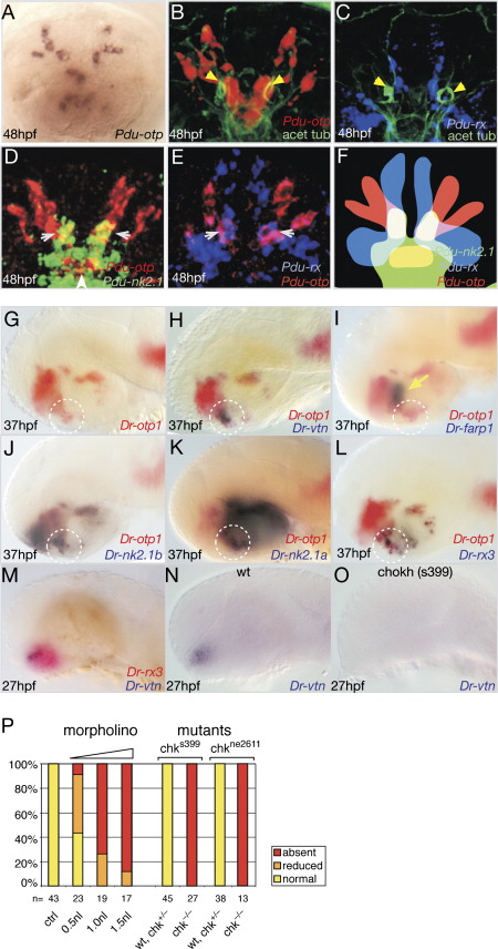
Fig. 6 Regulatory Signature of vtn+ and RFamidergic Cells and Dependence of Dr-vtn on Dr-rx3. Expression of indicated genes in (A–E) Platynereis and (G–O) zebrafish. (B and C) Expression in relation to the axonal scaffold (green), compare to Figures 3A–3C, 3J, 3L, and 5B for colocalization with Pdu-vtn, Pdu-c-opsin, miR-7. Yellow arrowheads: large cilia of deep brain ciliary photoreceptor cells. (F) Scheme of (D) and (E). The overlap of all three transcription factors (white) includes the medial vtn+ cells (white arrows in [D] and [E]). Overlap of nk2.1 and otp (yellow) in the medial RFamidergic cells (white arrowhead in [D]). (G–L) White circle demarcates same group of cells. Yellow arrow: Dr-farp1+ cells. (N and O) Absence of Dr-vtn expression in wild-type (WT) versus chks399 (chokh) siblings. (P) Summary of regulatory analyses, all assayed at 27–28 hpf. Morpholino-assisted knockdown of rx3 leads to dose-dependent reduction/absence of vtn expression at 27–28 hpf compared to controls (ctrl). Loss of vtn expression in early differentiating cells in two homozygous rx3 mutant alleles (chk s399 and chk ne2611). Below each bar: numbers of investigated specimens and volume of injected morpholino solution/genotype of fish as judged by presence/absence of eyes. Views: (A–F) apical, ventral to the bottom, (G–O) lateral, anterior left.
Reprinted from Cell, 129(7), Tessmar-Raible, K., Raible, F., Christodoulou, F., Guy, K., Rembold, M., Hausen, H., and Arendt, D., Conserved sensory-neurosecretory cell types in annelid and fish forebrain: insights into hypothalamus evolution, 1389-1400, Copyright (2007) with permission from Elsevier. Full text @ Cell