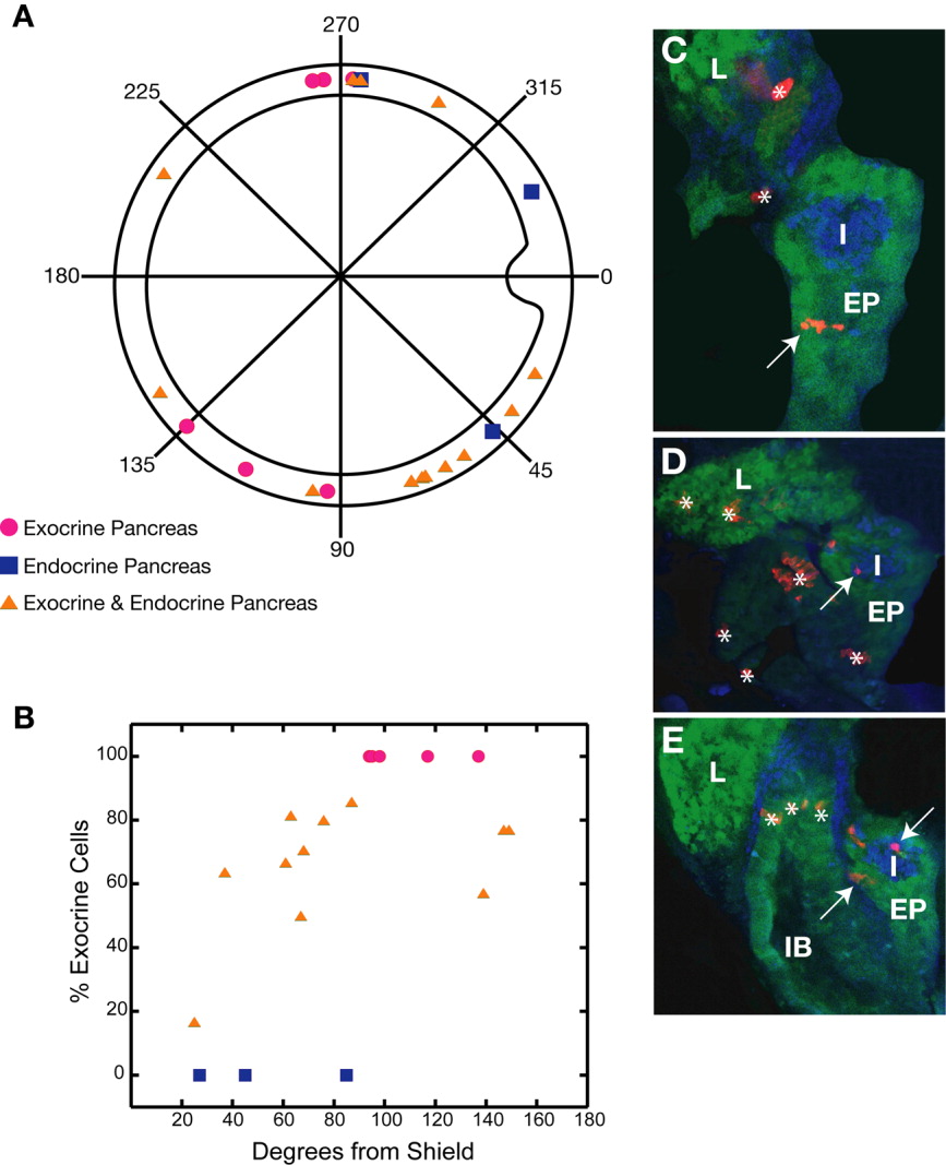
Fig. 4 Fate map of the endocrine and exocrine pancreas in zebrafish at 6 hours postfertilization (hpf). A: Polar plot of the locations of endocrine and exocrine pancreatic progenitors at mid-gastrulation. B: Graph of percentage exocrine cells at 72 hpf vs. dorsoventral location of the clone at 6 hpf. The numbers of exocrine and endocrine cells were counted, and the percentage exocrine is the number of exocrine cells divided by the total number of cells. C-E: Confocal slices of representative 72 hpf embryos. C: Embryo with rhodamine dextran-labeled cells in the exocrine pancreas. D: Embryo with labeled cells in the endocrine pancreas. E: Embryo with labeled cells in both the endocrine and exocrine pancreas. Blue cells are islet1 positive. Arrows point to labeled cells that colocalize with the structure of interest. Asterisks (*) point to rhodamine dextran-labeled cells that are not in the pancreas or liver. EP, exocrine pancreas; I, pancreatic islet; IB, intestinal bulb; L, liver.
Image
Figure Caption
Figure Data
Acknowledgments
This image is the copyrighted work of the attributed author or publisher, and
ZFIN has permission only to display this image to its users.
Additional permissions should be obtained from the applicable author or publisher of the image.
Full text @ Dev. Dyn.

