
Fig. 5
- ID
- ZDB-IMAGE-060424-6
- Genes
- Publication
- Grinblat et al., 1998 - Determination of the zebrafish forebrain: induction and patterning
- All Figures
- Figures for Grinblat et al., 1998
|
Fig. 5
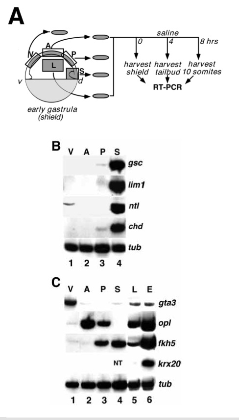
Forebrain pattern is specified at early gastrula. (A) Schematic outline of the explant assay. Four segments of early gastrula (shield stage) epiblast were isolated: ventral epiblast (V), anterior dorsal epiblast (A), posterior dorsal epiblast (P) and lateral epiblast (L). Explants of embryonic shield (S) contained a mixture of presumptive ectoderm, mesoderm and endoderm. All explants were combined in groups of 5 and harvested immediately (B) or cultured until control embryos reached tailbud or 10 somite (C) stage before analysis of marker gene expression by RT-PCR (see Methods). (B) RT-PCR analysis of explants harvested immediately after dissection and assayed for expression of dorsal mesendodermal markers gsc (Stachel et al., 1993; Thisse et al., 1994) and lim1 (Toyama et al., 1995), both markers of the axial hypoblast of the shield (prospective prechordal plate), ntl (Schulte-Merker et al., 1994), a marker of the entire mesendoderm, and chd (Miller-Bertoglio et al., 1997) a marker of the dorsal mesendodorm. α-tubulin (G. Conway, personal communication) was used as loading control. The data shown here is representative of 2 experiments. Lane 1: ventral explant; lane 2: anterior dorsal explant; lane 3: posterior dorsal explant; lane 4: shield explant. (C) RT-PCR analysis of cultured explants assayed for expression of ectodermal markers gta3 (Neave et al., 1995), a marker of ventral ectoderm, opl, a marker of prospective telencephalon, fkh5, a marker of prospective diencephalon, mesencephalon and spinal cord, krx20 (Oxtoby and Jowett, 1993), a marker of prospective rhombencephalon,and α-tubulin (G. Conway, personal comunication),aloading control. Each lane represents a pool of five explants. The data shown is representative of 3-4 independent experiments. Lane 1: ventral explants; lane 2: anterior dorsal explants; lane 3: posterior dorsal explants; lane 4: shield explants; lane 5: lateral ectoderm explants; lane 6: whole embryo control. NT: not tested.