
Fig. 6
- ID
- ZDB-IMAGE-050711-6
- Genes
- Publication
- Perkins et al., 2005 - dazed gene is necessary for late cell type development and retinal cell maintenance in the zebrafish retina
- All Figures
- Figures for Perkins et al., 2005
|
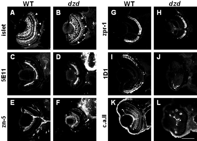
Fig. 6 Retinal cell type marker analysis of dazed mutants at 72 hours postfertilization (hpf). Central, transverse retinal sections of wild-type (WT, left) and dazed (right) were labeled with cell type-specific antibodies and visualized with appropriate secondary antibodies (see text for details). A,B: Islet-1 immunoreactivity; ganglion, bipolar, and horizontal cells and their precursors. C,D: 5E11 antigen; amacrine cells and their processes. E,F: zn-5 antigen; ganglion cells and their processes. G,H: zpr-1/fret-43 antigen; red-green double cones. I,J: 1D1 antigen; rod photoreceptors. K,L: Carbonic anhydrase II (c.a. ii) immunoreactivity; Müller glial cells and nonocular ectoderm. Scale bar = 80 μm in L (applies to A-L).