
Fig. 7
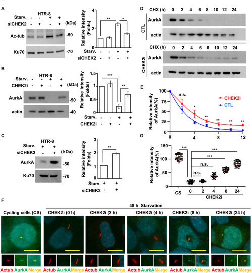
CHEK2 promotes AurkA degradation. A Depletion of CHEK2 reduced the amount of acetylated tubulin under serum starvation. Extracts of wild-type or CHEK2-depleted HTR-8 cells in the absence or presence of serum starvation (Starv.) were analyzed by Western blotting with antibodies against acetylated tubulin (Ac-tub), phosphorylated CHEK2 (p-CHEK2), and Ku70. B-C Inhibition or depletion of CHEK2 restores AurkA under serum deprivation. Extracts of HTR-8 cells treated with (B) CHEK2 inhibitor (CHEK2i) or (C) siRNA against CHEK2 (siCHEK2) were analyzed by Western blotting with antibodies against AurkA (AurA), Ku70, and actin. D-E Inhibition of CHEK2 increased AurkA protein stability. D Extracts of control (CTL) or CHEK2-inhibited (CHEK2i) cells with cycloheximide (CHX) treatment for 0, 2, 4, 6, 8, 10, 12, and 24 h were analyzed by Western blotting with antibodies against AurkA (AurA) and actin. E Quantitative results of D. F-G AurkA was reduced after 48 h of serum starvation and reappeared at the base of primary cilia. F AurkA in HTR-8 cells cultured in full medium (cycling cells) or serum-starved medium (48 h starvation) in the absence (CHEK2i 0 h) or presence of CHEK2i for different time periods (CHEK2i 2 h, 4 h, 8 h, and 24 h) was examined by immunofluorescence staining with antibodies against AurkA and acetylated tubulin (Actub). DNA were stained with DAPI. Scale bar: 10 μm. G Quantitative results for the relative intensity of AurkA in F. n.s.: no significance, *p < 0.05, **p < 0.01. These results are mean ± SD from three independent experiments |