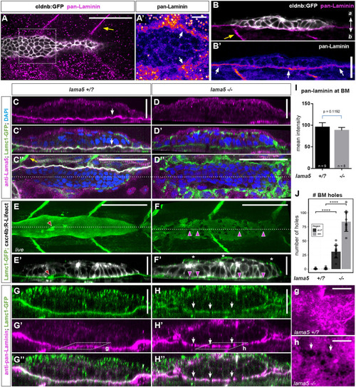
Laminin α5 is an integral component of the pLLP migration substrate. (A-B′) Pan-laminin immunostaining in whole-mount embryos (A,A′) and transverse cryosections (B,B′). Yellow and white arrows point to somite boundaries and the epidermal BM, respectively. Additional magnifications from A are in Fig. S8A-A″. (C-D″) Lama5 immunostaining in control (lama5+/?) (C-C″) and lama5−/− (D-D″) lamc1-GFP embryos. Orthogonal views (C-D′) and single z-planes (C″,D″) show Lama5 and Lamc1 in the epidermal BM (white arrows in C,C′) in controls. Lama5, but not Lamc1, is missing in the BM in lama5 mutants (D-D″). (E-F′) Live imaging of lamc1-GFP highlighting BM holes in lama5 mutants (purple arrowheads). (E-F′) Maximum-intensity projections of 5 μm substacks (E,F) and single orthogonal views (E′,F′). Lamc1-GFP also labels the Schwann cells of the PLLn (red arrowheads). (J) Quantification of the number of holes underneath pLLP (dark gray) and adjacent skin (light gray). (G-H″,g,h) Pan-laminin immunostaining in lamc1-GFP embryos. Orthogonal views (G-H″) and higher magnifications of single z-planes (g,h, as indicated in G′,H′), show similar discontinuities in the BM in lama5 mutants (white arrows in H-H″, h). (I) Quantification of pan-laminin signal intensity (data are mean±s.d.). (J) Dots represent data from individual embryo (data are mean±s.d.) Unpaired, two-tailed t-test; ****P<0.0001. Scale bars: 50 μm for top-views in A,C″,D″,E,F; 20 μm for optical sections in B,B′,C,C′,D,D′,E′,F′,G-H″; 10 μm for higher magnifications (A′,g,h). a, apical; b, basal.
|
