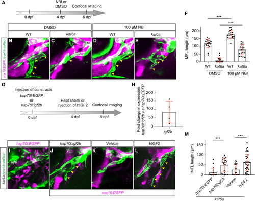
Enhancement of IGF signaling rescues the MFL defect in kat6ab719 mutants (A) Diagram illustrating the treatment regime for DMSO and the IGFBP inhibitor NBI-31772. (B–E) Confocal images of the lateral head in lyve1b:dsRed (green) sox10:EGFP (magenta) larvae at 6 dpf, comparing cartilage and lymphatic phenotypes between WT siblings (B) or kat6ab719 (C) treated at 4 dpf with DMSO and WT siblings (D) or kat6ab719 (E) treated at 4 dpf with 100 μM NBI. Yellow arrowheads highlight the MFL. (F) Quantification of the length of the MFL at 6 dpf, comparing the DMSO-treated WT (n = 20), DMSO-treated kat6ab719 (n = 18), NBI-treated WT (n = 21), and NBI-treated kat6ab719 (n = 20). (G) Diagram illustrating the workflow for the induction of Igf2b overexpression and injecting recombinant human IGF2. (H) RT-qPCR analysis of igf2b expression 2 h following heat shock (n = 4 experiments), confirming the overexpression of igf2b. (I–L) Confocal images of the lateral head in lyve1b:dsRed (green) sox10:EGFP (magenta) larvae at 6 dpf, comparing cartilage and lymphatic phenotypes between kat6ab719 larvae injected at one-cell stage with either the hsp70l:EGFP construct (I) or the hsp70l:igf2b construct (J) following a heat-shock treatment at 4 dpf at 37°C for 2 h and between kat6ab719 larvae injected at 4 dpf with either vehicle (K) or hIGF2 (L). Yellow arrowheads highlight the MFL. (M) Quantification of the length of the MFL at 6 dpf, comparing kat6ab719 injected with hsp70l:EGFP (n = 19), hsp70l:igf2b (n = 20), vehicle (n = 22), and 500 pg of hIGF2 (n = 35). MFL, medial facial lymphatic vessel. Data represent mean ± SD; ∗∗∗p < 0.001; ∗∗p < 0.01; ∗p < 0.05; ns, p > 0.05 from unpaired t test with Welch’s correction.
|
