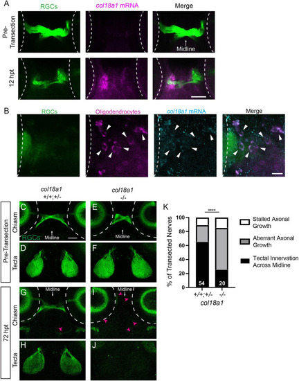
The Lh3 candidate substrate Col18a1 is upregulated after transection and directs RGC axons across the midline during regeneration. (A) col18a1 mRNA expression pre-transection and at 12 h post-transection (hpt) in zebrafish larvae expressing Tg(isl2b:GFP) to label RGCs. Axons in the chiasm region at 12 hpt have not yet fully degenerated following transection. Scale bar: 50 μm. (B) Additional images of individual oligodendrocytes labeled by Tg(olig2:dsred) co-expressing lh3 mRNA (see also Movie 4). Scale bar: 10 μm. (C-F) Before injury, RGC axons labeled by Tg(isl2b:GFP) in siblings (C,D) and col18a1 mutants (E,F) cross the midline and innervate contralateral tecta. (G-J) In contrast to col18a1 siblings at 72 hpt (G,H), RGC axons in col18a1 mutants turn away from the midline, project aberrantly (magenta arrowheads) and do not re-innervate contralateral optic tecta (I,J). Dashed lines indicate the outline of the eyes. Scale bar: 50 µm. (K) Quantification of the percentage of nerves at 72 hpt that grew across the midline to re-innervate the optic tectum, did not grow across the midline and instead grew along aberrant trajectories or showed stalled axonal growth. Numbers indicate total number of transected nerves. ****P<0.0001 (Fisher's exact test). Representative images of chiasms and tecta for each timepoint are of the same fixed larva, whereas across timepoints are of different larvae.
|
