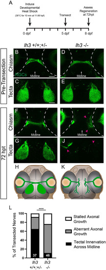
Lh3 is required for growth across the midline during regeneration. (A) Timeline of a transection experiment with zebrafish lh3 larvae (hpf, hours post-fertilization; dpf, days post-fertilization; hpt, hours post-transection). (B-E) Before injury, RGC axons labeled using Tg(isl2b:GFP) in siblings (B,C) and conditional lh3 mutants (D,E) cross the midline and innervate contralateral tecta. (F-H) In lh3 siblings at 72 hpt, RGC axons grow across the midline and re-innervate contralateral optic tecta, schematized in H. (I-K) In contrast, RGC axons in lh3 mutants turn away from the midline, project aberrantly (magenta arrowheads) and do not reinnervate contralateral optic tecta, schematized in K. Dashed lines indicate the outline of the eyes. Scale bars: 50 µm. Representative images of chiasms and tecta for each timepoint are of the same fixed larva, whereas across timepoints are different larvae. (L) Quantification of the percentage of nerves at 72 hpt that grew across the midline to re-innervate the optic tectum, did not grow across the midline and instead grew along aberrant trajectories, or showed stalled axonal growth. Numbers indicate total number of transected nerves. ****P<0.0001 (Fisher's exact test).
|
