
Fig. 3
- ID
- ZDB-FIG-251016-88
- Publication
- Park et al., 2025 - Lego Self-Assembly Model of Zebrafish RGB Cone Pentamer Formation: Unique Role of Crumbs2b in Cone Coalescence via Planar Orientational Cell Adhesions
- Other Figures
- All Figure Page
- Back to All Figure Page
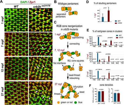
Crb2b deficiency gradually changes the isolated linear mirror-symmetric pentameric configuration of RGB cones into a bead–thread network. (A) Transverse imaging of RGB cones in the wild-type and crb2bsa21178 homozygous mutants of different ages. RGB cone nuclei were stained with DAPI (green), and green and red cones were stained with Zpr1 (red). Large white arrows indicate the groups of RGB cones depicted in panels B and C. Small double arrows indicate blue cones associated with only one cone square; arrowheads indicate square ends with no blue cones attached. (B) The diagram depicts the pentamers in the wild-type retina. (C) Diagrams depict the gradual alteration of the pentameric configuration into a bead–thread network pattern over time in crb2bsa21178 mutants. Orange indicates red or green cones, and green indicates blue cones. The bead–thread networks of RGB cones become apparent even at 7 mpf. Images were taken from the dorsal intermediate regions of the retina. (D) The percentages of abutting pentamers were higher in crb2bsa21178 mutants than those in wild-type retina at 1 mpf (n = 4 fish). (E) The percentages of green and red cones in clusters of various sizes. 1-C indicates isolated single cells; 2-C, two-cell clusters; etc. The percentages were compared between the wild-type and mutants at 7 mpf, 12 mpf, and 27 mpf (n = 4 fish). (F) The densities of RGB and UV cones in the wild-type and crb2bsa21178 homozygous mutants at 27 mpf (n = 4 fish; one tangential confocal image per fish). The P values were calculated by Student's t-test. |