
Fig. 1
- ID
- ZDB-FIG-251016-86
- Publication
- Park et al., 2025 - Lego Self-Assembly Model of Zebrafish RGB Cone Pentamer Formation: Unique Role of Crumbs2b in Cone Coalescence via Planar Orientational Cell Adhesions
- Other Figures
- All Figure Page
- Back to All Figure Page
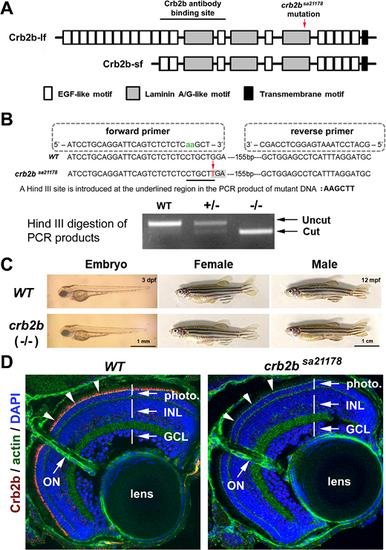
crb2bsa21178 mutation eliminates Crb2b expression in the retina with no gross defects. (A) The position of crb2bsa21178 mutation and the Zou anti-Crb2b antibody binding site in the extracellular domains of the Crb2b-lf and Crb2b-sf. The EGF-like, laminin A/G-like transmembrane motifs are illustrated as shaded boxes. (B) The strategy to genotype wild-type (WT) and crb2bsa21178 mutant (–/–) fish by PCR and Hind III digestion. (C) crb2bsa21178 mutant fish showed no apparent differences in body shape at 3 dpf and adulthood compared to the wild-type. (D) Crb2b immunostaining signals (red, arrowheads) were eliminated in crb2bsa21178 homozygous mutant retina at 5 dpf. The photoreceptor layer (photo), inner nuclear layer (INL), ganglion cell layer (GCL), lens, and optic nerve (ON) were revealed by phalloidin staining of actin and DAPI staining of the cell nuclei. |