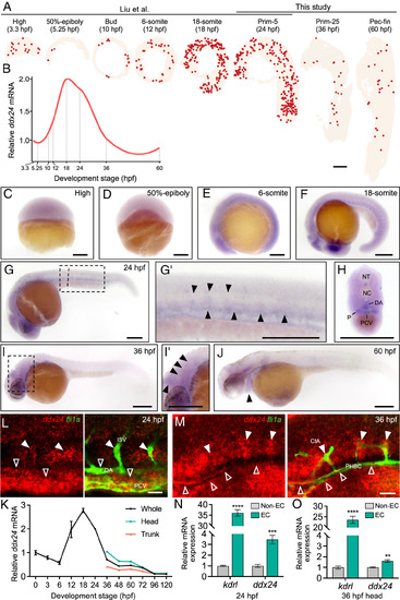
Expression pattern of ddx24 in developing zebrafish. (A) Spatial visualization of ddx24 expression across zebrafish developmental stages. Data for 3.3 to 24 hpf are from Liu et al., while data for 24 to 60 hpf are from this study. (B) Relative mean expression of ddx24 across all cell types at various developmental stages. (C–J) Whole-mount in situ hybridization (WISH) showing ddx24 mRNA expression at various developmental stages. (C–F) Early embryos exhibit ubiquitous ddx24 expression. (G) Lateral view of 24 hpf embryos showing ddx24 mRNA expressed in the head and trunk. (G′) Magnified view of the trunk region from (G), highlighting enriched ddx24 expression in the vasculature. Arrowheads indicate the trunk vessels. (H) Transverse sections of the embryo trunk from (G) showing the vascular expression of ddx24. DA, dorsal aorta; PCV, posterior cardinal vein; NT, neural tube; NC, notochord; P, pronephric duct. (I and J) At 36 and 60 hpf, ddx24 expression is restricted to the brain and heart regions. (I′), Magnified view of the head region in (I). Arrowheads indicate the brain in (I′) and the heart in (J). (K), Quantitative real-time PCR (qRT-PCR) analysis of ddx24 mRNA expression in whole embryos, as well as in the head and trunk, during development. (L and M), Fluorescence WISH for ddx24 (red) combined with anti-EGFP staining (green) in Tg(fli1a:EGFP)y1 embryos. Colocalization of ddx24 and EGFP in the trunk vasculature at 24 hpf (L) and brain vasculature at 36 hpf (M). ISV, intersegmental vessels; PHBC, primordial hindbrain channels; CtA, central artery. In (L), the solid arrow indicates ISVs, while the hollow arrow indicates the DA and PCV. In (M), the solid arrow indicates tip cells of CtAs, while the hollow arrow indicates PHBC. (N and O), qRT-PCR of ddx24 mRNA in both GFP+ and GFP- cells from Tg(fli1a:EGFP)y1 transgenic embryos at 24 and 36 hpf. kdrl mRNA serves as a positive control for vascular endothelial cells. [Scale bars, 200 μm in (A and C–J), 20 μm in (L and M).] Data are represented as mean ± SD. **P < 0.01, ***P < 0.001 and ****P < 0.0001, as assessed by parametric two-tailed Student’s t test.
|

