
Fig. 3
- ID
- ZDB-FIG-250521-8
- Publication
- Xu et al., 2025 - Microglia-Derived IL-6 Promotes Müller Glia Reprogramming and Proliferation in Zebrafish Retina Regeneration
- Other Figures
- All Figure Page
- Back to All Figure Page
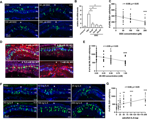
IL-6 promotes MGPC formation in the injured zebrafish retina. (A) EdU immunofluorescence showing the cell proliferation in PBS control or Di-O-demethylcurcumin (DDC)-treated retinas at 4 dpi. (B) The qPCR analysis of il6 mRNA levels in the PBS- or DDC-treated retinas at 12 hpi. Uninjured retina (0 day) served as a negative control. (C) Quantification of the number of INL EdU+ cells per injury at 4 dpi of A. (D) EdU immunofluorescence showing the cell proliferation in retinas electroporated with lissamine-tagged control (ctrl) MO or il6r MO at 4 dpi. (E) Quantification of the number of INL EdU+ cells per injury at 4 dpi of D. (F) EdU immunofluorescence showing the cell proliferation in retinas treated with PBS control or indicated doses of zebrafish IL-6 at 4 dpi. (G) Quantification of the number of INL EdU+ cells per injury in PBS or IL-6-treated retinas at 4 dpi. White *, site of the stab injury. *, P < 0.05; **, P < 0.01; ***, P <0.001. GCL, ganglion cell layer; INL, inner nuclear layer; ONL, outer nuclear layer; r, correlation coefficient. |