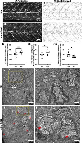
Loss of mtmr5 leads to less defined axonal organization and dysregulated axon myelination. (A and B) Seven days post-fertilization (dpf) zebrafish embryos were fixed and probed with an acetylated tubulin antibody to label mature axons. (A and B) Representative images of Z-projections (standard deviation via Fiji ImageJ) of acetylated tubulin staining. Axons appear less organized in the mtmr5-knockout (KO) compared to the sibling. (Ai and Bi) Two-dimensional skeletonized images generated using Binary function in Fiji ImageJ to allow for quantification of axonal organizational parameters. (C-Cii) The number of total axonal branches (C) and axon end-points (Ci) significantly increased (by ∼25%) in the KO compared to the siblings (Sib), while the average axon branch length (Cii) in KOs significantly decreased (by ∼10%). All quantification was normalized to the sibling average of the respective parameter. All statistical analyses include at least three independent experiments. Each dot on the graphs represents one zebrafish. Quantitative data are mean±SEM, normalized to the average of WT and presented as ratios. Unpaired two-tailed Student’s t-test was used. *P < 0.05; **P < 0.01, ns, not significant. Scale bars (A and B): 20 μm. Sib n = 11, KO n = 10. (D) Transmission electron microscopy was used to visualize axons in the posterior lateral line at 14 dpf. WT axons were in close contact with their myelin sheaths. This tight and organized wrapping (D, box) can be observed in more detail in the zoomed-in inset (Di). SC = Schwann cell. (E) In the 14 dpf KO, the myelin is still present; yet, there is an apparent blank space between the axon and the myelin (arrows), showing the detachment or loose myelin in the KOs (E, yellow box; Ei). Scale bars: (D and E) 2 μm or (Di and Ei) 0.5 μm.
|
