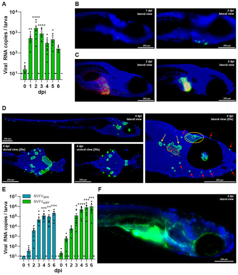
Viral replication and dissemination of RVFVeGFP in zebrafish larvae, with infection of the liver, sensory nervous system and vascular system. (A) Viral RNA copies of RVFVeGFP per zebrafish larva. Mean ± s.e.m. of 6–8 pools of 10 larvae from 8 independent experiments. (B–D,F) Whole mount immunohistochemistry fluorescence images of RVFVeGFP-infected zebrafish larvae at 10× magnification, using anti-GFP primary antibody and Hoechst 33342. (B) Wildtype zebrafish larva at 1 and 2 dpi showing progressing RVFV infection around the site of injection. (C) Tg (fabp10a:DsRed; nacre) zebrafish larvae demonstrating infection in the liver using anti-RFP primary antibody. (D) Wildtype zebrafish larva showing infection of the retina, optic tectum (yellow circle), the anterior and posterior macula (red circle), the ganglia (yellow arrows), and neuromasts (red arrows) of the lateral line. dpi = days post infection. (E) Viral RNA copies of RVFV35/74 and RVFVeGFP per zebrafish larva immersed in Danieau’s solution supplemented with ruxolitinib (25 µM) from 0 dpi. Inoculum: ~1 CCID50 of RVFV35/74 or ~15 CCID50 of RVFVeGFP. Mean ± s.e.m. of 8 pools of 10 larvae from 8 independent experiments. (F) Wildtype zebrafish larva at 6 dpi, showing extensive dissemination of RVFVeGFP infection after immersion in Danieau’s solution supplemented with ruxolitinib (25 µM) from 0 dpi. Image has not been 3D-deconvoluted. dpi = days post infection; RVFV = Rift Valley fever virus; * p < 0.05; ** p < 0.01; *** p < 0.001; **** p < 0.0001; ns = not significant.
|
