
Fig. S1
- ID
- ZDB-FIG-241009-8
- Publication
- Ugur et al., 2024 - VPS13B is localized at the interface between Golgi cisternae and is a functional partner of FAM177A1
- Other Figures
- All Figure Page
- Back to All Figure Page
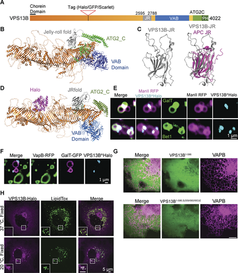
Structure and localization of VPS13B. (A) Domain structure of human VPS13B. (B) Alphafold2 predicted structure of full-length human VPS13B; orange indicates the rod consisting of 13 RBG domains. (C) Alphafold2 prediction of the Jelly Roll (JR) domain of VPS13B alone on the left and overlayed with the jelly-roll fold of anaphase-promoting complex subunit Doc1p/Apc10 on the right. (D) Alphafold2 prediction of VPS13B internally tagged with Halo tag (magenta). (E) Snapshots of Golgi complex fragments of HeLa cells expressing VPS13B^Halo, ManII-RFP (a medial-Golgi marker), and either GalT-GFP (top panel) or Bet1-GFP (bottom panel) after a 10-min hypotonic shock. Scale bar = 1 µm. (F) Snapshots of Golgi complex and ER fragments of HeLa cells expressing VPS13B^Halo, VapB-RFP, and GalT-GFP after a 10-min hypotonic shock. Scale bar = 1 µm. (G) Top panel: HeLa cells co-expressing VapB-RFP and the N-terminal fragment of VPS13B (a.a. 1–586). Bottom panel: HeLa cells co-expressing VapB-RFP and the mutant N-terminal fragment (a.a. 1–586): VPS13BS559E, S560E, S563E. Scale bar = 10 µm. (H) COS7 cells expressing VPS13B^Halo and kept at 37°C (top panel) or shifted to 20°C for 30 min before fixation (bottom panel) and then stained with Lipid Tox. Scale bar = 5 µm. |