
Fig. 3
- ID
- ZDB-FIG-241009-11
- Publication
- Ugur et al., 2024 - VPS13B is localized at the interface between Golgi cisternae and is a functional partner of FAM177A1
- Other Figures
- All Figure Page
- Back to All Figure Page
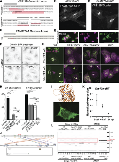
Generation of VPS13B and FAM177A1 KO HeLa cells and evidence that the zebrafish genome encodes and expresses a VPS13B homolog. (A) Sanger sequencing of VPS13BKO1 and VPS13BKO2 HeLa cells; superscripts indicate different clones. (B)VPS13BKO2 HeLa cells expressing FAM177A1-GFP. (C) Sanger sequencing of FAM177A1KO homozygous HeLa cells. (D)FAM177A1KO cells expressing VPS13B^Scarlet. Scale bar = 10 µm. (E) Snapshots of Golgi fragments of a VPS13BKO2 HeLa cell expressing ZFPL1-GFP, and GalT-RFP after a 10-min hypotonic shock. Scale bar = 5 µm. (F) Anti-GM130 immunofluorescence of WT, VPS13BKO1, FAM177A1KO, and VPS13B;FAM177A1 DKO cells after 30 min in BFA (5 µg/ml). (G) WT, VPS13BKO1, FAM177A1KO, and FAM177A1;VPS13B DKO HeLa cells transfected with ZFPL1-GFP and ManII-RFP after 1 h in BFA (5 µg/ml) followed by subsequent washings as indicated. (H) Quantification of Golgi complex reformation in cells of the indicated genotypes after BFA washout for 2 or 5 h. Data are mean ± SEM n = 3 per condition; in each condition, 20–50 cells were quantified. Unpaired, two-tailed t tests. n.s., not significant. ***P < 0.001; **P < 0.01; *P < 0.05. (I) Top: Sites (blue) within the RBG structure of VPS13B where hydrophobic amino acids facing the floor of the hydrophobic grove were replaced by charge amino acids (L65K, I81E, L90E, I155R, L169E, A176E, I203R, L238D, I355K, L264R) to generate the Lipid Transport Dead (LTD) Mut1. Bottom: HeLa cell expressing VPS13LTDmut1 showing small clusters of the protein sparse throughout the cytoplasm rather than a Golgi localization. (J) Conserved syntenies of VPS13B in human and zebrafish validate orthology implied by sequence comparisons. A small part of human (Homo sapiens) chromosome 8 (Hsa8, green part in insert) has conserved synteny with a short portion of zebrafish (Danio rerio) chromosome16 near its right tip (Dre16, green portion in insert). (K) qRT-PCR analysis of WT vps13b in early zebrafish embryos at two-cell stage, 6 hpf, 24 hpf. (L) Genotyping of vps13b CRISPR target locus in WT zebrafish embryos injected with vps13b gRNAs/Cas9 and treated with 0.8 µg/ml BFA (samples 1–6), fam177a1a;fam177a1b DKO embryos injected with vps13b gRNAs/Cas9 and treated with 0.8 µg/ml BFA (samples 7–12), WT zebrafish embryos injected with vps13b gRNAs/Cas9 and treated with DMSO (samples 13–18), WT zebrafish embryos treated with DMSO (samples 19, 20), and fam177a1a;fam177a1b DKO embryos injected with vps13b gRNAs/Cas9 and treated with DMSO (samples 21, 22). |