
FIGURE
Fig. 3
- ID
- ZDB-FIG-240419-61
- Publication
- Paulissen et al., 2024 - Live Imaging Transverse Sections of Zebrafish Embryo Explants
- Other Figures
- All Figure Page
- Back to All Figure Page
Fig. 3
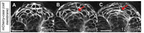
Time-lapse imaging of cell polarization in the neural keel. A. Image of the neural keel starting at the 10-somite stage. B. Image of the neural keel after 60 min. Note the polarization of cells at the midline (red arrow). C. Image of neural keel after 120 min observation during the transition to the neural rod. Note the cells have polarized at the midline (red arrow ). Scale bars = 50 μm. |
Expression Data
Expression Detail
Antibody Labeling
Phenotype Data
Phenotype Detail
Acknowledgments
This image is the copyrighted work of the attributed author or publisher, and
ZFIN has permission only to display this image to its users.
Additional permissions should be obtained from the applicable author or publisher of the image.
Full text @ Bio Protoc