
Fig. 6
- ID
- ZDB-FIG-240314-6
- Publication
- Shin et al., 2022 - leptin b and its regeneration enhancer illustrate the regenerative features of zebrafish hearts
- Other Figures
- All Figure Page
- Back to All Figure Page
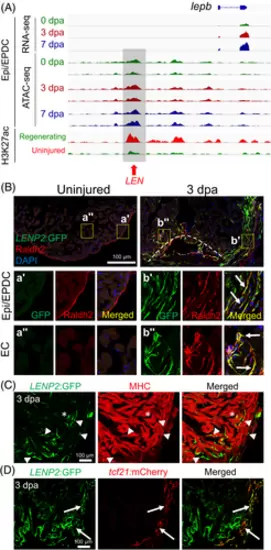
LEN directs injury-induced Epi/EPDC expression. (A) Genome browser tracks of the genomic region near lepb showing the transcripts and chromatin accessibility profiles in the Epi/EPDC. The whole-ventricle H3K27Ac profile of the uninjured and regenerating heart is shown at the bottom. Gray box and red arrow indicate LEN. (B-D) Immunostained section images of transgenic fish carrying LENP2:EGFP. (B) Raldh2 antibody is used to label EC and Epi/EPDC. Uninjured heart shows one single Raldh2+ cell layer outlining the cardiac chamber. Raldh2 signal emerges in the ECs at the wound area and EPDCs in the cortical layers upon injury, which are co-labelled with LENP2:EGFP (Arrows). The boxed areas are enlarged at the bottom panels. (C) Myosin heavy chain (MHC) antibody is used to label CMs. (D) tcf21:mCherry is used for Epi/EPDC expression. While LENP2:EGFP rarely colocalizes with MHC+ CMs (Arrowheads), a subset of LENP2:EGFP co-localizes with tcf21:mCherry (Arrows). Note that asterix indicates CM expression as a basal expression of the P2 minimal promoter. At least five hearts for uninjured and injured samples were examined and all animals displayed a similar expression pattern |