FIGURE

Figure 2.

ID
ZDB-FIG-240213-2
Publication
Kahsay et al., 2024 - Obscurin Maintains Myofiber Identity in Extraocular Muscles
Other Figures
All Figure Page
Back to All Figure Page
Figure 2.

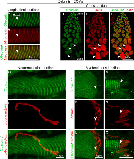
Obscurin distribution in zebrafish EOMs. (A–C) Longitudinal sections of 10-month-old wild-type zebrafish EOMs immunolabeled with antibodies against obscurin (green in A), and myomesin (red in B; merged image in C). Obscurin is localized in the M-band identified by staining with antibodies against myomesin (red in B). (D–F) Immunolabeling against obscurin (green in D) and F-actin (red in E; merged in F) showed the presence of obscurin in the vast majority of the myofibers in zebrafish EOMs. Lack of obscurin was also noted in a small subgroup of myofibers (arrowheads). Longitudinal sections of zebrafish EOMs immunolabeled with obscurin (green in G) at NMJs identified by α-bungarotoxin (red in H; merged in I) showed that obscurin was not enriched at NMJs as in other muscles. Immunolabeling of obscurin (arrowheads, green in J, M) at MTJs of zebrafish EOMs labeled by laminin (red in K, N; merged in L and O).

Expression Data

Expression Detail
Antibody Labeling
Phenotype Data

Phenotype Detail
Acknowledgments
This image is the copyrighted work of the attributed author or publisher, and ZFIN has permission only to display this image to its users. Additional permissions should be obtained from the applicable author or publisher of the image. Full text @ Invest. Ophthalmol. Vis. Sci.