FIGURE SUMMARY
Title

Obscurin Maintains Myofiber Identity in Extraocular Muscles

Authors
Kahsay, A., Dennhag, N., Liu, J.X., Nord, H., Rönnbäck, H., Thorell, A.E., von Hofsten, J., Pedrosa Domellöf, F.
Source
Full text @ Invest. Ophthalmol. Vis. Sci.

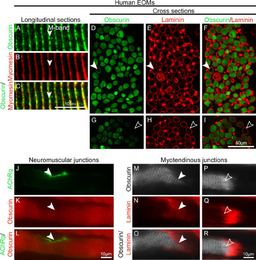
Localization and distribution of obscurin in human EOMs. (A–C) Longitudinal sections of human EOMs immunolabeled with antibodies against obscurin (green in A) and myomesin (red in B; merged in C), arrowheads indicate the M band. (D–I) Adjacent cross-sections of human EOM immunostained with antibodies against obscurin (green in D and G) and laminin (red in E and H; merged images in F and I). Note that obscurin was present in the vast majority of the human EOM myofibers. In a small percentage of myofibers, obscurin was present only subsarcolemally (arrowheads in D–F) or absent (open arrowheads in G–I). (J–R) Longitudinal sections of human EOM are shown. Obscurin (red in K and L) was maintained at the NMJs labeled by ACHRγ (arrowheads, green in J and L) rather than to be enriched, as known from the limb and trunk muscles.19 (M–R) Obscurin was also maintained (arrowheads, gray in M and O) in the majority of MTJs, but it was increased (open arrowheads, gray in P and R) at some MTJs. MTJs of human EOMs were identified by their localization and typical labeling by laminin (arrowheads, red in N and O, Q and R).

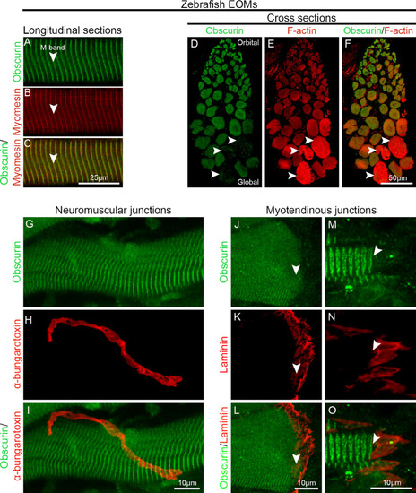
Obscurin distribution in zebrafish EOMs. (A–C) Longitudinal sections of 10-month-old wild-type zebrafish EOMs immunolabeled with antibodies against obscurin (green in A), and myomesin (red in B; merged image in C). Obscurin is localized in the M-band identified by staining with antibodies against myomesin (red in B). (D–F) Immunolabeling against obscurin (green in D) and F-actin (red in E; merged in F) showed the presence of obscurin in the vast majority of the myofibers in zebrafish EOMs. Lack of obscurin was also noted in a small subgroup of myofibers (arrowheads). Longitudinal sections of zebrafish EOMs immunolabeled with obscurin (green in G) at NMJs identified by α-bungarotoxin (red in H; merged in I) showed that obscurin was not enriched at NMJs as in other muscles. Immunolabeling of obscurin (arrowheads, green in J, M) at MTJs of zebrafish EOMs labeled by laminin (red in K, N; merged in L and O).

Obscna and obscnb knockout zebrafish (A) Schematic representation of generation of knockouts using CRISPR/Cas9 gene editing technology in zebrafish. (B, C) Amino acid sequence illustrating Obscna and Obscnb protein. Blue letters indicate the frameshift mutation resulting in premature stop codon indicated by asterisk. The resulting genotyped PCR products after cutting with restriction enzymes (obscna; BbVCI; New England BioLabs, Ipswich, MA, USA) and obscnb; SacI (Thermo Scientific, Vilnius, Lithuania) are shown using gel electrophoresis. DNA sequences of obscna and obscnb knockouts are displayed with chromatography, and mutation sites are shown in the box. (D) Obscurin immunolabeling of obscna+/;obscnb+/ and obscna/;obscnb/ zebrafish larvae at 5dpf. Longitudinal sections of 10-month-old zebrafish EOMs immunolabeled with (E) α-bungarotoxin labeling multiple nerve endings (arrowheads) in obscna+/;obscnb+/ and obscna/;obscnb/ adult zebrafish. (F) F-actin (green) and Tenascin (red) immunolabeling in whole EOMs of 10-month-old zebrafish showing MTJs of myofibers of obscna+/;obscnb+/ and obscna/;obscnb/ adult zebrafish. (G) myomesin (green) labeling M-band, desmin (red) labeling Z-discs (desmin-positive myofiber on the left column, and desmin-negative myofiber on the right column). (H) Cross-sections of 10-month-old zebrafish EOMs: subsarcolemmal localization of desmin in obscna/;obscnb/ and distributed throughout the myofiber in obscna+/;obscnb+/ (arrowheads indicate sarcolemma, open arrowheads indicate cytoplasm). (I) Percentage of myofibers showing desmin distribution either subsarcolemmaly or throughout the myofiber relative to the total number of myofibers. ****P < 0.0001.

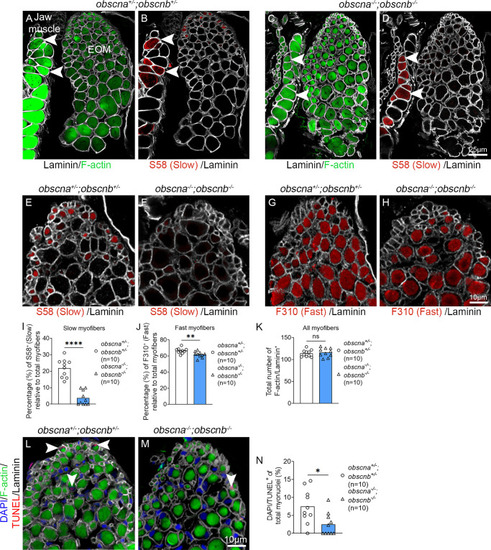
Quantification of slow and fast myofibers in the EOMs of adult obscurin mutants and sibling controls. Cross-sections of 10-month-old adult zebrafish EOMs immunolabeled with phalloidin to identify all myofibers (labeling of F-actin by phalloidin,
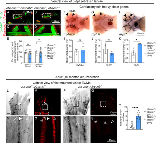
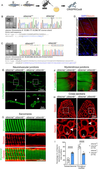
 green in A and C), S58 to identify all myofibers containing slow MyHC (red in B, D–F), F310 to identify all myofibers containing fast MyHC (red in G and H). The contours of the myofibers were labeled by the antibody against laminin (gray in A–H). Arrowheads indicate examples of positively-labeled myofibers with phalloidin, which labels F-actin (in A and C) and slow MyHC S58 (in B and D) in closely located masticatory myofibers (left) in the same section. Quantification of slow myofibers (S58 positive in E and F and quantified in I), fast myofibers (F310 positive in G and H and quantified in J), and the total number of myofibers (K) in obscna/;obscnb/ and obscna+/;obscnb+/. (L, M) Cross-sections of obscna+/;obscnb+/ and obscna/;obscnb/ zebrafish EOMs immunolabeled with phalloidin, which labels F-actin, laminin, DAPI and TUNEL (arrowheads, red in L and M) to identify apoptotic myofibers. (N) The proportion of TUNEL-positive myofibers. Data in graphs are presented as mean ± SEM. *P < 0.05; **P < 0.01; ****P < 0.0001.

Expression of myh7 myofibers of zebrafish EOMs. (A–D) Ventral view of the zebrafish EOMs (dashed squares) in double transgenic lines, Tg(mylz2:GFP, green, identifies all fast myofibers) and Tg(smyhc:tdTomato, red, identifies all slow myofibers) of obscna+/;obscnb+/ and obscna/;obscnb/ zebrafish larvae at 5 dpf. C and D show the EOMs in the marked areas in A and B at higher magnification. (E) Measurement of fast (identified by Tg(mylz2:GFP) and slow Tg(smyhc:tdTomato) myofibers total size (µm) in the EOMs relative to controls measured in midportion of the EOMs, indicated by white line in C, presented in percentage. (F–H) Ventral view of wild-type zebrafish larvae at 5dpf showing expression of cardiac myosin heavy chain genes, (F) myosin heavy chain 7bb (myh7bb), (G) myosin heavy chain 7-like(myh7l), and (H) myosin heavy chain 7 (myh7) probes in the EOMs and the cardiac muscle, arrowheads indicate EOMs, MM = masticatory muscle and CM = cardiac muscle. The qPCR showed the mRNA level of (I) myh7bb, (J) myh7l, and (K) myh7 in obscna/;obscnb/ and obscna+/;obscnb+/ zebrafish larvae. Expression of myh7 was significantly increased in EOMs. (L–O) EOMs from obscna+/;obscnb+/ treated with myh7 antisense probe showing small subgroup of myh7-positive myofibers (L, N, arrowheads), never overlapping with S58 labeled slow myofibers (M, O, open arrowheads) and (P–S) EOMs from obscna/;obscnb/ where open arrowheads indicate myh7-positive myofibers. The areas indicated by the squares in L, M, P, and Q are shown below in higher magnification. (T) Quantification of the number of myh7 positive myofibers in obscna/;obscnb/ (n = 9) and obscna+/;obscnb+/ (n = 11). *P < 0.05; ****P < 0.0001.

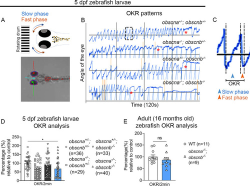
Optokinetic response analysis of the zebrafish EOMs: (A) Schematic representation of slow phase (blue) and fast phase (red) of the zebrafish eye. (B) Representative OKR patterns of obscna+/;obscnb+/, obscna/;obscnb+/, obscna+/;obscnb/ and obscna/;obscnb/ over 120 seconds. (C) A magnified image of WT OKR pattern showing slow and fast phases representing one OKR count. (D) Percentage of OKRs relative to control in 5 dpf zebrafish larvae. (E) Percentage of OKRs relative to controls in 16-month-old adult zebrafish.

Acknowledgments
This image is the copyrighted work of the attributed author or publisher, and ZFIN has permission only to display this image to its users. Additional permissions should be obtained from the applicable author or publisher of the image. Full text @ Invest. Ophthalmol. Vis. Sci.