
Fig. 1
- ID
- ZDB-FIG-231227-30
- Publication
- Greysson-Wong et al., 2023 - rasa1-related arteriovenous malformation is driven by aberrant venous signalling
- Other Figures
- All Figure Page
- Back to All Figure Page
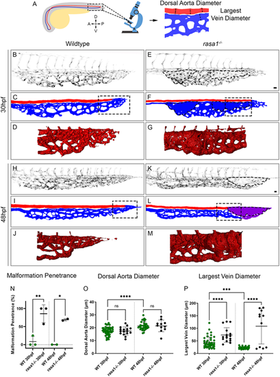
rasa1 mutants show vessel enlargement in the caudal venous plexus without affecting the dorsal aorta. (A) Schematic of the location where confocal images were taken of the caudal venous plexus (outlined). Schematic of how the dorsal aorta (DA) and largest vein diameter were measured. A, anterior; P, posterior; D, dorsal; V, ventral. (B,E,H,K) Confocal images of the caudal venous plexus of wild-type and rasa1 mutant endothelium (kdrl:EGFP, black). (C,F,I,L) Schematic of the vessels showing the dorsal aorta (red), caudal venous plexus (blue) and vessels of unknown arteriovenous identity (purple). (D,G,J,M) Simpleware was used on high-resolution confocal images to create 3D renderings of the flow return area where malformations develop in rasa1 mutants. Areas outlined in C,F,I,L indicate the approximate location of the 3D renderings from images of different embryos. (N-P) Quantification of confocal images of wild-type and rasa1 mutant embryos at 30 hpf and 48 hpf. (N) Penetrance of vessel enlargement (≥1.5× average largest wild-type vein diameter) at 30 hpf and 48 hpf (30 hpf: wild type, n=13; rasa1−/−, n=16; N=3 experiments, P=0.005; 48 hpf: wild type, n=10; rasa1−/−, n=10; N=2 experiments, P=0.024). (O) The average wild-type DA diameter was not significantly different from that in mutants at either timepoint (30 hpf: wild type, n=13, rasa1−/−, n=16; N=3 experiments, P>0.99; 48 hpf: wild type, n=12; rasa1−/−, n=11; N=3 experiments, P>0.99). (P) The largest vein at 30 hpf is larger in mutants and is further enlarged at 48 hpf (30 hpf: wild type, n=13; rasa1−/−, n=16; N=3 experiments, P=0.0018; 48 hpf: wild type, n=12; rasa1−/−, n=11; N=3 experiments, P<0.0001). Statistical analysis used a one-way ANOVA with Sidak's correction. Data are mean±s.d. Scale bars: 20 µm. |