
Fig. 4
- ID
- ZDB-FIG-231215-88
- Publication
- Chen et al., 2020 - The mevalonate pathway is a critical regulator of tendon cell specification
- Other Figures
- All Figure Page
- Back to All Figure Page
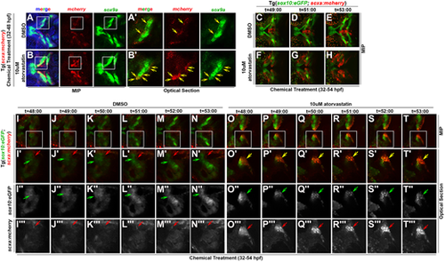
Statin expands the scxa-expressing cell population by promoting tenogenic specification of CNC. (A-B′) Expression of mcherry and sox9a in (A,A′) DMSO and (B,B′) atorvastatin-treated Tg(scxa:mcherry) embryos at 48 hpf. White boxes mark the craniofacial domain in maximum intensity projection (MIP) (A,B), magnified in corresponding optical sections (A′,B′). Yellow arrows mark mcherry+/sox9a+ cells. Atorvastatin expanded mcherry+/sox9a+ cells (B′, compare with A′). (C-T‴) Time-course of live imaging of tendon and cartilage development in (C-E,I-N‴) DMSO- and (F-H,O-T‴) atorvastatin-treated Tg(sox10:eGFP;scxa:mcherry) embryos (n=4 shown). Arrows mark sox10:eGFP+ cells of Meckel's cartilage (green), scxa:mcherry+ tendon progenitors (red) and scxa:mcherry+/sox10:eGFP+ cells (yellow). Atorvastatin expanded scxa:mcherry+/sox10:eGFP+ cells near the region where Meckel's cartilage forms (compare F-H and O-T‴ with C-E and I-N‴, respectively). White boxes mark the craniofacial domain in the MIPs (I-T) of high-resolution images, magnified in the corresponding MIPs (I′-T′) and optical sections: eGFP (I″-T″) and mcherry (I‴-T‴) expression. Ventral views, anterior towards the left. |