
Fig. 4
- ID
- ZDB-FIG-231215-19
- Publication
- Edwards et al., 2023 - Hemato-vascular specification requires arnt1 and arnt2 genes in zebrafish embryos
- Other Figures
- All Figure Page
- Back to All Figure Page
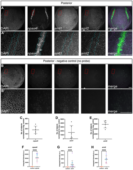
arnt1 and arnt2 are ubiquitously expressed at the 4-somite stage and colocalized with npas4l expression. Whole-mount in situ hybridization chain reaction was performed on wild-type (AB) embryos at the 4-somite stage. (A) Embryos were probed for npas4l, arnt1 and arnt2 expression, and counterstained with DAPI. arnt1 and arnt2 labeling was detected ubiquitously, while npas4l labeling was localized to the lateral plate mesoderm. (A′) Higher magnification views of the areas outlined in A shows colocalization of arnt1, arnt2 and npas4l in the posterior lateral plate mesoderm, outlined in cyan. (B) To demonstrate that the observed expression of arnt1 and arnt2 is not autofluorescence of the embryo or the yolk, wild-type embryos were processed identically to those in A, but no antisense RNA probe sets were added. (B′) Higher magnification views of the areas outlined in B shows what background/autofluorescence looks like in the presumptive posterior lateral plate mesoderm. All images in A and B are maximum intensity projections from z-stacks taken every 2.5 μm. In merged panels, npas4l is green, arnt1 is magenta and arnt2 is cyan. The posterior region of the embryo is in view, dorsal is towards the top of the image. Scale bars: 100 μm. (C-E) To validate the observed expression of arnt1 and arnt2, we quantified the number of detectable objects above a set threshold in the whole embryo across each z-plane for four negative control embryos and six experimental embryos probed for arnt1, arnt2 and npas4l. We then normalized the number of detected objects by subtracting the average number of objects detected in the negative controls from the number of detected objects in the experimental embryos. Each point on these graphs represents the total number of objects above the threshold for a single embryo. (F-H) To demonstrate that arnt1 and arnt2 expression occurs in the lateral plate mesoderm (LPM), we calculated the integrated intensity of fluorescence (pixel area and pixel intensity) over each side of the posterior LPM in maximum intensity projections from control and experimental embryos. Each point represents the integrated intensity from one side of the posterior LPM from a single embryo (right or left side), where each embryo has two bilateral npas4l-positive regions of the posterior LPM. Points of the same color are measurements from opposite sides of the posterior LPM from the same embryo. npas4l, arnt1 and arnt2 labeling was statistically significantly increased in the lateral plate mesoderm compared with negative control embryos. Data are mean±s.e.m. npas4l Welch's t-test, t(11)=8.6, ****P<0.0001; arnt1 Mann–Whitney, U=0, ****P<0.0001; arnt2 Welch's t-test, t(11)=7.0, ****P<0.0001. |