
Fig. 7
- ID
- ZDB-FIG-231102-29
- Publication
- Zhao et al., 2023 - Metaphocytes are IL-22BP-producing cells regulated by ETS transcription factor Spic and essential for zebrafish barrier immunity
- Other Figures
- All Figure Page
- Back to All Figure Page
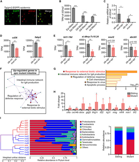
Metaphocytes are the major source of Il22ra2 and modulate immunity in barrier tissues (A) ISH detection of il22ra2 in the epidermis of TgBAC(grn2:EGFP) fish. Scale bar, 50 μm. (B) TPM values of il22ra2 in metaphocytes and macrophages of zebrafish barrier tissues by bulk RNA-seq. Values are represented as mean ± SD (n ≥ 7). ∗∗∗p < 0.001. Epi, epidermis; Int, intestine; Meta, metaphocytes; Macro, macrophages. (C) qPCR detection of the relative expression level of il22ra2 in the epidermis and intestine of spicΔ136 mutants and siblings. eef1a1l1 was utilized as the internal control. Values are represented as mean ± SD (n = 4). ∗p < 0.05. epi, epidermis; int, intestine; sib, sibling; mut, mutant. (D) TPM values of lipid transporter and metabolic genes in the intestine of spicΔ136 mutants and siblings by whole-tissue RNA-seq. Values are represented as mean ± SD (n = 4). ∗∗p < 0.01, ∗∗∗p < 0.001. (E) TPM values of keratinocyte terminal differentiation marker genes in the epidermis of spicΔ136 mutants and siblings by whole-tissue RNA-seq. Values are represented as mean ± SD (n = 4). ∗p < 0.05, ∗∗p < 0.01. (F) Main GO clusters enriched in the upregulated genes of spicΔ136 mutant intestine. (G) GO terms enriched in the upregulated genes of spicΔ136 mutant intestine. (H) Expression fold change of the representative genes of the GO term “response to external biotic stimulus” in the intestine of spicΔ136 mutants compared with that in siblings. Values are represented as mean ± SD (n = 4). ∗p < 0.05, ∗∗p < 0.01. (I) Left: cluster tree of spicΔ136 mutants and siblings based on intestinal microbiome composition by UPGMA method. Right: the relative abundance of intestinal microbiome phylum in adult spicΔ136 mutants and siblings. See also Figure S5. |