|
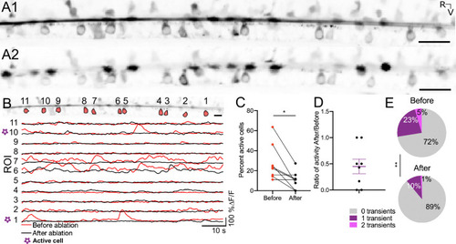
The Reissner fiber enhances spontaneous calcium activity in ventral CSF-cNs. (A1) Time-series standard deviation projection in the sagittal plane from two-photon laser scanning microscope showing the signal from the Reissner fiber and CSF-cNs in the central canal of 3 dpf Tg(sspo:sspo-GFP;pkd2l1:tagRFP;pkd2l1:GCaMP5G) zebrafish larva before RF photoablation. (A2) Time-series standard deviation projection in the sagittal plane from two-photon laser scanning microscope showing the signal from CSF-cNs after RF ablation performed by spiral scanning photoablation with an infrared pulsed laser tuned at 800 nm over 0.5 μm on RF (see Materials and methods). (B) ROI selection for ventral CSF-cNs to analyze activity before and after RF photoablation within the same cells (top). Example calcium activity traces normalized to baseline for each of the ROIs before (red) and after (black) RF photoablation over 75 s imaged at 3.45 Hz (see Materials and methods). (C) Percentage of active ventral CSF-cNs (active is defined as having at least 1 calcium transient during the recording) before and after RF photoablation (N=109 cells total from 8 fish from two independent clutches; mean percent active before ablation = 27.72% ± 6.34% versus mean percent active after ablation = 10.59% ± 3.1%; paired two-tailed t-test: p < 0.05). (D) The ratio of active ventral CSF-cNs after RF photoablation to those active before RF photoablation, illustrating on average, a fraction (on average ± SEM: 45% ± 14%) of active ventral CSF-cNs before photoablation remain active after RF photoablation. The purple lines on the graph represent the mean and the error bars indicate the SEM. (E) Pie charts illustrating the number of events per ventral CSF-cN before and after RF photoablation (mean number of events in active cells before RF photoablation = 0.94 events/min versus mean number of events in active cells after RF photoablation = 0.87 events/min; paired two-tailed t-test: p < 0.005). * p < 0.05, **p < 0.005. Scale bar is 20 μm (A1, A2), 10 μm (B).
|

