|
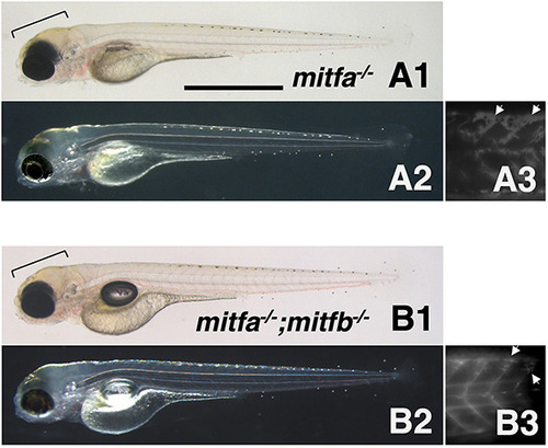
Phenotypes of zebrafish mitfa and mitfb double homozygotes. (A1-B3) 4 dpf mitfa−/− (nacre) hatchling (A1-A3) and 4 dpf mitfa−/−; mitfb−/− hatchling (B1-B3) under normal transmission optics (A1,B1), dark-field epi-illumination optics (A2,B2) and autofluorescence images showing xanthophores under UV light (A3,B3). Yellowish-pigmented xanthophores are observed in the dorsal head of the mitfa−/− hatchling (A1,A2). Autofluorescence emitted by xanthophores is clearly visible in the trunk of mitfa−/− (A3). Similarly, the mitfa−/−; mitfb−/− hatchling has xanthophores in the head (B1,B2) and autofluorescent cells in the trunk (B3). Brackets indicate pigmented xanthophores and arrows indicate autofluorescent (possibly immature) xanthophores. Scale bar: 1 mm.
|