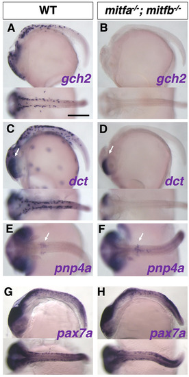
In situ analyses of medaka mitfa−/−; mitfb−/− double mutants with pigment cell markers. Results of in situ analyses with specification markers, dct for melanophore, gch2 for xanthophore/leucophore, and pnp4a for iridophore, are consistent with the phenotypes of differentiated pigment cells. The gch2-expressing xanthophore/leucophore progenitors are lost in the double mutant (A,B). The dct-expressing melanophore progenitors are absent from the body surface, but not from the eyes (retinal pigment epithelium, RPE) in the double-mutant embryo (C,D; arrows indicate the signal in RPE), consistent with the phenotype that the mutant retains melanized RPE (see Fig. 8K1,K3). The pnp4a-expressing iridophore progenitors appear unchanged in the double mutant compared with WT (E,F; arrows indicate the signal on the yolk). The mitfa−/−; mitfb−/− double mutant shows normal expression pattern of pax7a mRNA compared with WT (G,H), suggesting that the defect in xanthophore and leucophore formation in the double mutant is not mediated by Pax7a function. Lateral views at top and dorsal views at bottom. (A-D,G,H) Stage 28. (E,F) Stage 29. Scale bar: 250 µm.
|
