
Fig. 2
- ID
- ZDB-FIG-231031-42
- Publication
- Matsui et al., 2023 - Phosphorylation of α-synuclein at T64 results in distinct oligomers and exerts toxicity in models of Parkinson's disease
- Other Figures
- All Figure Page
- Back to All Figure Page
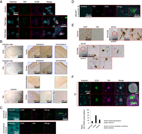
Distinct pathology of T64-phosphorylated α-synuclein. (A) T64-P and S129-P α-synuclein immunofluorescence of the SH-SY5Y cells transfected with α-synuclein fibrils. α-Synuclein Aggregation Assay Kit was used for the transfection of α-synuclein fibrils and cells are subjected to immunofluorescence at 1 and 3 d after transfection. (B) Immunohistochemistry of T64-P and S129-P α-synuclein in the mouse PD models (α-synuclein PFF mouse). α-Synuclein fibrils were injected into the striatum of C57BL/6J mice at 5 mo. Two months after inoculation, the mice were sacrificed and the brains were subjected to immunohistochemistry. (C) Immunofluorescence of T64-P and S129-P α-synuclein in the mouse PD models (α-synuclein PFF mouse). α-Synuclein fibrils were injected into the striatum of C57BL/6J mice at 5 mo. Two months after inoculation, the mice were killed, and the brains were subjected to immunofluorescence. (D) Immunofluorescence of T64-P and S129-P α-synuclein in the zebrafish PD models. Zebrafish (Tg(XenopusNBT:human α-synuclein);gba-/-) were killed and subjected to immunofluorescence at 3 mo of age. (E) Immunohistochemistry of T64-P and S129-P α-synuclein in the human PD brains. The medulla oblongata sections of human postmortem brains were used for immunohistochemistry. Patient profiles are described in SI Appendix, Table S3. (F) Immunofluorescence of T64-P and S129-P α-synuclein in the human PD brains. The medulla oblongata sections of human postmortem brains were used for immunofluorescence. Patient profiles are described in SI Appendix, Table S3. |