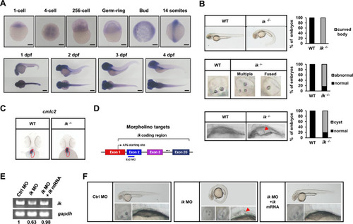
zebrafish ik mutants display ciliopathy-like phenotypes (A) Whole-mount in situ hybridization (WISH) analysis of zebrafish ik mRNA at different developmental stages (1-cell, 4-cell, 256-cell, Germ-ring, Bud, 14 somites, 1 dpf, 2 dpf, 3 dpf, and 4 dpf). Scale bar, 250 μm (B) Brightfield microscopic images of the body curvature, otolith, and pronephric cysts pronephros in WT embryos and ik mutants (ik−/−) at 2 dpf. Pronephric cyst of ik mutants is marked with red arrowhead. Stacked bar graph displays the percentage of embryos with a ventrally curved body, abnormal otolith phenotype, and cyst formation (C) WISH analysis of cmlc2 for the location of whole heart in WT embryos and ik mutants at 2 dpf (D) Schematic representation of the coding region of zebrafish ik. The target region (exon 2) of the morpholino employed in this study was represented using a blue line (E) RT-PCR analysis of ik mRNA expression in control morphants (MO), ik MO, and ik MO/ik mRNA co-injected embryos at 2 dpf. gapdh served as a normalization control and band intensity ratio of ik/gapdh mRNA expression was marked (F) Brightfield microscopic images of control MO, ik MO, and ik MO/ik mRNA co-injected embryos at 2 dpf. Whole body, otolith, and kidney cyst were captured. Pronephric cysts of ik MO are marked with red arrowhead
|
