FIGURE

Fig. 1

ID
ZDB-FIG-231002-96
Publication
Anderson et al., 2023 - Ligament injury in adult zebrafish triggers ECM remodeling and cell dedifferentiation for scar-free regeneration
Other Figures
All Figure Page
Back to All Figure Page
Fig. 1

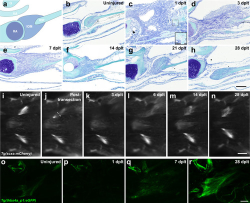
Adult zebrafish ligament transection injury and regeneration.

a Schematic of the articulating bones at the zebrafish jaw joint and connections of the interopercular-mandibular (IOM) ligament. bh Toluidine blue staining of adult zebrafish IOM ligament in uninjured (b, n = 16), 1 day post-ligament transection (dplt, c, n = 3), 3 dplt (d, n = 5), 7 dplt (e, n = 6), 14 dplt (f, n = 16), 21 dplt (g, n = 3), and 28 dplt (h, n = 16). in Repeated live imaging of the ventral view of scxa:mCherry expression in IOM ligament before injury (i, n = 9), immediately after injury (j, n = 3), 3 dplt (k, n = 9), 6 dplt (l, n = 3), 14 dplt (m, n = 9), and 28 dplt (n, n = 3). o–r Confocal microscopy imaging of tissue cleared thbs4a_p1:GFP expression in IOM ligament uninjured control (o, n = 6), 1 dplt (p, n = 5), 7 dplt (q, n = 6), and 28 dplt (r, n = 3). AA, anguloarticular bone; IOM, interopercular-mandibular ligament; IOP, interopercular bone; Q, quadrate bone; RA, retroarticular bone. Scale bars = 100 μm (bh, or), 250 μm (in).

Expression Data

Expression Detail
Antibody Labeling
Phenotype Data

Phenotype Detail
Acknowledgments
This image is the copyrighted work of the attributed author or publisher, and ZFIN has permission only to display this image to its users. Additional permissions should be obtained from the applicable author or publisher of the image. Full text @ NPJ Regen Med