
Fig. 6
- ID
- ZDB-FIG-230905-10
- Publication
- Mishra-Gorur et al., 2023 - Pleiotropic role of TRAF7 in skull-base meningiomas and congenital heart disease
- Other Figures
- All Figure Page
- Back to All Figure Page
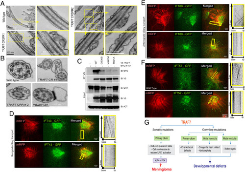
TRAF7 interacts with IFT57, and its knockdown in X. tropicalis impairs intraflagellar transport. (A and B). TEM micrographs of cilia from the pronephros of WT- and TRAF7-morphant Xenopus embryos at 5 dpf. Low- and high (yellow boxes)-magnification views of flat-embedded sections of longitudinally sectioned cilia show frequent blebbing only in TRAF7 morphant samples. Cross-sections of cilia from WT and morphant embryos show the “9 + 2” microtubule doublet configuration with the presence of dynein arms. Sections of cilia in TRAF7 morphant embryos show frequent blebs containing electron-dense material. (C and D). In vivo imaging of IFT dynamics in Xenopus multiciliated cells. Still frames from a video of IFT43-GFP (C) and IFT80-GFP (D) to track intraflagellar transport in a multiciliated cell (Movies S14–16). Cilia are colabeled with mRFP. The yellow box indicates the cilia shown in the right kymograph, depicting still frames from a time-lapse video showing movement of a single control cilium. Time is indicated in seconds. (Scale bars, 10 µm.) (E) Meningioma and CHD- and craniofacial defect-associated TRAF7 mutants (G536S, V442M, and T601A, respectively) show reduced interaction with IFT57. Coimmunoprecipitation analysis in HEK293 cells. (F) In vivo imaging of IFT dynamics in Xenopus multiciliated cells. Still frames from a video of IFT57-GFP to track intraflagellar transport in a multiciliated cell (Movies S17 and S18). Cilia are colabeled with mRFP. The yellow box indicates the cilia shown in the right kymograph, depicting still frames from a time-lapse video showing movement of a single control cilium. Time is indicated in seconds. (Scale bars, 10 µm.) (G) TRAF7 mutations disrupt ciliogenesis resulting in developmental (congenital heart and craniofacial) defects and disease (anterior skull-base meningioma). |