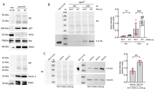
The E262K mutant of δ-SG is the substrate of the ERAD in zebrafish. (A) Western blot analysis of the expression of different components of the endoplasmic reticulum associated degradation (ERAD) in zebrafish. Lysates from 5 dpf larvae of the wild type zebrafish were resolved in SDS-PAGE and probed with antibodies specific for the indicated proteins (representative WBs are reported). The antibodies used are listed in Table S2; M, molecular markers; PR, Ponceau red staining used for protein content normalization. (B) sgcd−/− zebrafish oocytes were microinjected with two concentrations (as indicated) of the human mRNA encoding either the wild type or mutated E262K-δ-SG. Embryos at 48 hpf were lysed and the total proteins purified. A representative WB (left), and densitometric quantification of human-δ-SG bands (right) are shown. The graph reports the band intensity values of the E262K-δ-SG (MUT) and wild type (WT) forms of the human protein at different concentrations of injected mRNA. The mean values ± SD from at least four independent microinjection experiments are reported. Statistical analysis was performed by one-way ANOVA followed by Šídák’s multiple comparisons test; ns, p > 0.05; ***, p ≤ 0.001. (C) sgcd−/− zebrafish oocytes, at one cell stage, were microinjected with 1200 pg of the human E262K-δ-SG mRNA. At 24 hpf, embryos were treated with DMSO 1‰ (vehicle) or MG132 (10 µM). At 56 hpf, the embryos were lysed and the total proteins purified. A representative WB (left) and the densitometric quantification of human-δ-SG bands (right) are shown. The graph reports the band intensity values of the mutated protein in the two groups of embryos. The mean values ± SD from five independent microinjection experiments are reported. PR, Ponceau red staining. Blots were probed with rabbit polyclonal antibodies specifically recognizing the human δ-SG or tubulin, used for protein content normalization. Proteins purified from the HEK293 cells transfected with the human δ-SG sequence are reported as the positive control. Statistical analysis was performed by the Mann–Whitney test; **, p ≤ 0.01.
|
