
Fig. 7
- ID
- ZDB-FIG-230717-132
- Publication
- Nakajima et al., 2023 - Endoderm-derived islet1-expressing cells differentiate into endothelial cells to function as the vascular HSPC niche in zebrafish
- Other Figures
- All Figure Page
- Back to All Figure Page
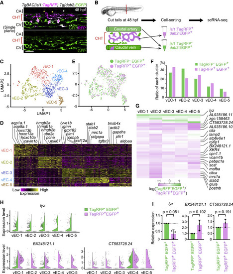
Figure 7. Transcriptomic heterogeneity and specialization in isl1-derived venous ECs constituting the vascular HSPC niche (A) Single confocal plane of the tail of a TgBAC(isl1:TagRFP);Tg(dab2:EGFP) embryo (48 hpf). Isl1:TagRFP+/dab2:EGFP+ cells are mostly present in venous ECs of the CHT (arrows). Isl1:TagRFP expression is also detected in the vISVs (yellow arrowheads) and motor neurons (white arrowhead). (B) Experimental design for scRNA-seq from the tail of TgBAC(isl1:TagRFP);Tg(dab2:EGFP) embryos. (C) Uniform manifold approximation and projection (UMAP) visualization of tail venous ECs corresponding to the venous EC cluster in Figure S6. Reclustered cells are color-coded for the 5 identified sub-clusters. (D) Heatmap illustrating the expression of the top-ranking marker genes enriched in each sub-cluster. Representative gene names are shown on top. (E) Mapping of isl1:TagRFP−/dab2:EGFP+ cells (772 cells: green) and isl1:TagRFP+/dab2:EGFP+ cells (449 cells: pink) into the UMAP shown in (C). (F) Percentage contribution of isl1:TagRFP−/dab2:EGFP+ cells and isl1:TagRFP+/dab2:EGFP+ cells to each venous EC sub-cluster. (G) Heatmap illustrating the log-count expression, log2(gene expression in isl1:TagRFP− EGFP+/gene expression in isl1:TagRFP+ dab2:EGFP+), of the 36 differentially expressed genes between isl1:TagRFP−/dab2:EGFP+ cells and isl1:TagRFP+/dab2:EGFP+ cells (q value < 0.05 in at least one sub-cluster). (H) Violin plots of the representative result of three genes; lyz, BX248121.1, and CT583728.24 in (G). The expression of lyz is dominant in isl1:TagRFP−/dab2:EGFP+ over isl1:TagRFP+ dab2:EGFP+ cells, whereas that of BX248121.1 and CT583728.24 is dominant in isl1:TagRFP+ dab2:EGFP+ cells over isl1:TagRFP−/dab2:EGFP+ cells. (I) isl1:TagRFP−/dab2:EGFP+ cells and isl1:TagRFP+/dab2:EGFP+ cells were isolated from the tail of TgBAC(isl1:TagRFP);Tg(dab2:EGFP) embryos at 48 hpf by fluorescence-activated cell sorting (FACS). Relative expression of lyz mRNA, BX248121.1 mRNA, and CT583728.24 mRNA in FACS-sorted cells was analyzed by quantitative PCR. Data are normalized to the values in isl1:TagRFP−/dab2:EGFP+ cells (fold change = 2(−ΔΔCt)). Data are mean ± SD. Each dot represents an individual experiment (n = 3 independent experiments). Scale bars, 50 μm. See also Figure S6 and Tables S1 and S2. |
Reprinted from Developmental Cell, 58(3), Nakajima, H., Ishikawa, H., Yamamoto, T., Chiba, A., Fukui, H., Sako, K., Fukumoto, M., Mattonet, K., Kwon, H.B., Hui, S.P., Dobreva, G.D., Kikuchi, K., Helker, C.S.M., Stainier, D.Y.R., Mochizuki, N., Endoderm-derived islet1-expressing cells differentiate into endothelial cells to function as the vascular HSPC niche in zebrafish, 224-238.e7, Copyright (2023) with permission from Elsevier. Full text @ Dev. Cell