
FIGURE
Fig. 1
Fig. 1
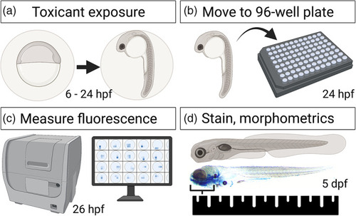
Experimental timeline. (a) Embryos were exposed from 6 to 24 hpf in petri dishes. (b) At 24 hpf, embryos were manually removed from their chorions and loaded into individual wells of a 96‐well plates. (c) At 26 hpf, embryos were scanned and imaged in vivo on a plate reader. (d) Embryos were then developed to 5 dpf, upon which time embryos were euthanized, fixed, stained, imaged, and finally morphologically measured. hpf = hours post fertilization, dpf = days post fertilization |
Expression Data
Expression Detail
Antibody Labeling
Phenotype Data
Phenotype Detail
Acknowledgments
This image is the copyrighted work of the attributed author or publisher, and
ZFIN has permission only to display this image to its users.
Additional permissions should be obtained from the applicable author or publisher of the image.
Full text @ Birth Defects Res