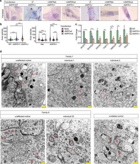
Patient-derived fibroblasts with AMFR loss-of-function variants show disturbed lipid metabolism and altered endoplasmic reticulum morphology a Representative images of ORO staining identifying lipids in unrelated wild type, heterozygous carrier (unaffected mothers from Family 1 and 8) and homozygous AMFR patient-derived fibroblasts from individuals 1, 2, and 20, and in the same fibroblasts transfected with plasmids expressing wild-type AMFR or RING mutant AMFR. Scale bars = 10 µm. b Left violin plots showing the size area of the quantified lipid droplets in pixels from ORO stained fibroblasts from a). Data from 2 wild-type fibroblasts (green colored), 2 heterozygous carrier AMFR fibroblasts (unaffected mothers of Family 1 and 8, purple colored) and 3 homozygous AMFR patient-derived fibroblasts (from individuals 1, 2, and 20, orange colored) (biological replicates), cultured and stained each in two technical replicates, assessing n ≥ 400 droplets for each sample. Black circle, median; black line, SD (Dunn’s Multiple Comparison test, ***p < 0.001). Right violin plot shows the same quantification from a), but then for heterozygous or homozygous AMFR fibroblasts transfected with plasmids expressing wild type AMFR or RING mutant AMFR, as indicated. c qRT-PCR expression analysis of genes involved in cholesterol metabolism in wild type (n = 2), heterozygous (n = 2, from unaffected mothers of Family 1 and 8), and homozygous (n = 3, from individuals 1, 2, and 20) AMFR patient-derived fibroblasts. Bar plot showing the mean fold change for the indicated genes compared to wild type, normalized for the housekeeping gene TBP. Each fibroblast line was cultured in two independent duplicates, and measured using two technical replicates and two independent experiments. Error bars represent SEM (Dunn’s Multiple Comparison test, *p < 0.05; **p < 0.01; ***p < 0.001). d Electron microscopy of cultured fibroblasts from individuals 1, 2, and 20, harboring homozygous truncating AMFR variants, as well as their respective heterozygous carrier mothers and an unrelated control. Affected individuals show an abundance of large vesicles indicative of lipid droplets as well as large, dilated rough endoplasmic reticulum (RER, red asterisks) as compared to the more compact RER seen in unaffected parents and an unrelated control (red + signs). Scale bars = 500 nm
|
