
Fig. 2
- ID
- ZDB-FIG-230706-21
- Publication
- van der Stoel et al., 2022 - Vinculin strengthens the endothelial barrier during vascular development
- Other Figures
- All Figure Page
- Back to All Figure Page
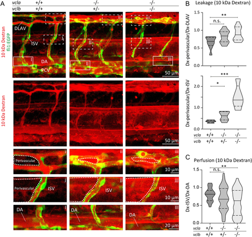
Vinculin ensures vascular barrier function for 10 kDa dextran. (A) Images of ISVs from 48 hpf Tg(fli1:EGFP) vcla+/+;vclb+/+, vcla−/−;vclb+/− or vcla−/−;vclb−/− embryos injected with 10 kDa rhodamine-dextran (red). Lower panels are single-channel images of the rhodamine signal. Scale bars, 50 µm. (A, i-iii) Corresponding close-up images showing the dextran leakage in the perivascular area around the dorsal longitudinal anastomotic vessel (DLAV). (A, j-jjj) Corresponding close-up images showing the dextran leakage in the perivascular area around an intersegmental vessel (ISV). Scale bars, 10 µm. (A, l-lll) Close-up images of an ISV and the dorsal aorta (DA) showing the rhodamine signal within these vessels. Scale bar, 20 µm. (B) Violin plots showing the average leakage ± s.e. of 10 kDa dextran into the perivascular area of the DLAV or ISVs normalised to the dextran inside the DLAV or ISVs from 48 hpf embryos. The dotted lines represent the quartiles and the straight lines represent the median, n = 10 vcla+/+;vclb+/+, n = 25 vcla−/−;vclb+/− and n = 13 vcla−/−;vclb−/− embryos, n.s., non-significant, *P < 0.05, **P < 0.01, ***P < 0.001 (one-way ANOVA and Dunnett’s post-test). PCV, posterior cardinal vein. (C) Violin plot showing the ISV perfusion determined as the ratio between fluorescent dextran levels inside the ISV ± s.e. and dextran inside the dorsal aorta (DA) from 48 hpf embryos. The dotted lines represent the quartiles, the straight lines represent the median. n = 10 vcla+/+;vclb+/+, n = 25 vcla−/−;vclb+/− and n = 13 vcla−/−;vclb−/− embryos. |