FIGURE

Figure 4

ID
ZDB-FIG-230630-28
Publication
Stemerdink et al., 2023 - Generation and Characterization of a Zebrafish Model for ADGRV1-Associated Retinal Dysfunction Using CRISPR/Cas9 Genome Editing Technology
Other Figures
All Figure Page
Back to All Figure Page
Figure 4

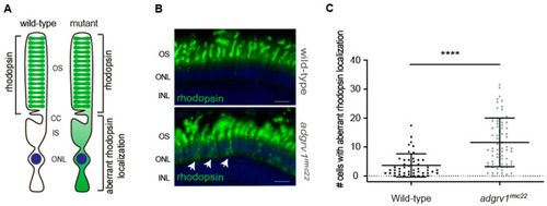
Aberrant localization of rhodopsin in photoreceptor cell bodies in the adgrv1rmc22 zebrafish. (A): Schematic representation of a photoreceptor with rhodopsin localization in the outer segments (OS) versus aberrant rhodopsin localization in the photoreceptor cell body as observed in adgrv1rmc22 mutants. (B): Retinal cryosections of wild-type and adgrv1rmc22 zebrafish larvae (6 dpf) labeled with antibodies directed against rhodopsin (green). Nuclei are counterstained with DAPI (blue). A significantly higher number of photoreceptors with aberrant localization of rhodopsin was observed in adgrv1rmc22 larvae (indicated with the white arrows) than in wild-type larvae. (C): Total number of cells with aberrant rhodopsin localization per retinal section were plotted, with mean ± SD (n = 29 adgrv1rmc22 mutant larvae and n = 21 wild-type larvae). A two-tailed unpaired Student’s t-test revealed a significant difference between adgrv1rmc22 mutants and wild-types. **** indicates p < 0.0001, scale bar: 10 μm. CC: connecting cilium; IS: inner segment; ONL: outer nuclear layer; INL: inner nuclear layer.

Expression Data

Expression Detail
Antibody Labeling
Phenotype Data

Phenotype Detail
Acknowledgments
This image is the copyrighted work of the attributed author or publisher, and ZFIN has permission only to display this image to its users. Additional permissions should be obtained from the applicable author or publisher of the image. Full text @ Cells