
Figure 5
- ID
- ZDB-FIG-230615-10
- Publication
- Schellens et al., 2023 - A protein domain-oriented approach to expand the opportunities of therapeutic exon skipping for USH2A-associated retinitis pigmentosa
- Other Figures
- All Figure Page
- Back to All Figure Page
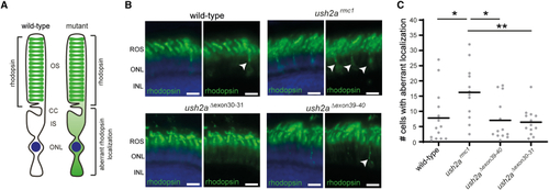
Visualization of rhodopsin in retinal sections of wild-type, ush2armc1, ush2aΔexon30-31, and ush2aΔexon39-40 zebrafish (A) In the photoreceptors of wild-type zebrafish, rhodopsin predominantly localizes to the outer segments, whereas in photoreceptors of mutant zebrafish, rhodopsin is defectively localized. (B) Retinal cryosections of larvae (6 dpf) were analyzed for rhodopsin (green) localization. Nuclei were counterstained with DAPI (blue). In the retinas of wild-type, ush2aΔexon30-31, and ush2aΔexon39-40 larvae, rhodopsin was predominantly present in the photoreceptor outer segments, whereas in ush2armc1 retinas rhodopsin signal was also detected in the photoreceptor cell bodies as indicated by arrowheads. Scale bar, 10 μm. INL, inner nuclear layer; ONL, outer nuclear layer; ROS, rod outer segment. (C) Scatterplot of detected photoreceptor cells with aberrant rhodopsin localization in 6 dpf zebrafish obtained by manual counting and plotted per counted eye (n = 12–14). Horizonal bars depict the mean number of photoreceptor cells with aberrant rhodopsin localization within a genotype. The number of cells with aberrant rhodopsin localization is significantly lower in wild-type, ush2aΔexon30-31, and ush2a |