|
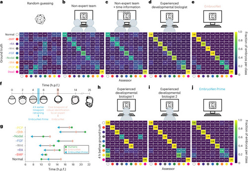
Classification of 98 single embryo images by non-expert teams, experienced researchers and EmbryoNet a–e, Schematic set-ups and confusion matrices showing the classification of the respective labeler compared with the ground truth (human annotation, treatment known). Classification performance is shown as a heatmap and fractions of 1 for the classification of 98 single images by a pseudo-random number generator (a), by a non-expert team without (b) or with (c) additionally provided time information (average performance), by an experienced researcher (d) and by EmbryoNet (e). f, Schematic of embryo detection over time. To allow for earlier detection, we annotated the training data 4 h before (blue time frame) the timepoint at which they could be robustly annotated by a labeler aware of the treatment (pink time frame). The embryo sketches show the phenotype of Nodal-inhibited samples at the respective time. The resulting network with earlier detection was termed ‘EmbryoNet-Prime’. g, Characteristic times of detection for each class based on the assessment of human experts, EmbryoNet and EmbryoNet-Prime. nNormal = 74, n−BMP = 119, n+RA = 66, n−Wnt = 70, n−FGF = 74, n−Nodal = 110, n−Shh = 63, n−PCP = 57. h–j, Classification performance in the early detection of phenotypes. Confusion matrices show the classification of image series by the respective labeler compared with the ground truth (human annotation, treatment known; detection time shifted to 4 h earlier). The number of analyzed images is shown in Supplementary Tables 20–22.
|