
FIGURE
Figure 5
- ID
- ZDB-FIG-230526-31
- Publication
- White et al., 2023 - The Presence of Two MyoD Genes in a Subset of Acanthopterygii Fish Is Associated with a Polyserine Insert in MyoD1
- Other Figures
- All Figure Page
- Back to All Figure Page
Figure 5
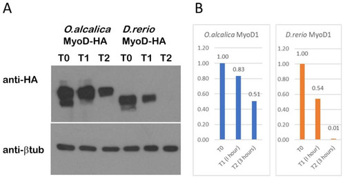
Stability assay shows more perdurance of O.alcalica MyoD1 compared to D. rerio MyoD1. (A) Western blot analysis of extracts from Xenopus laevis explants overexpressing HA-tagged MyoD1 proteins. Explants were incubated for either 0 (T0), 1 (T1) or 3 h (T2) in the translational inhibitor, cycloheximide. Anti- beta tubulin is a loading control. (B) Quantification of the loss of protein relative to the amount of protein detected at T0 is shown in a graph for O.alcalica MyoD1 and for D. rerio MyoD1. |
Expression Data
Expression Detail
Antibody Labeling
Phenotype Data
Phenotype Detail
Acknowledgments
This image is the copyrighted work of the attributed author or publisher, and
ZFIN has permission only to display this image to its users.
Additional permissions should be obtained from the applicable author or publisher of the image.
Full text @ J Dev Biol