
Figure 1
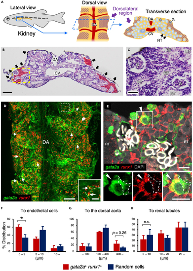
gata2a+runx1+ HSPCs are closely associated with vascular endothelial cells in the kidney (A) Schematic diagram of zebrafish kidney. Lateral view (left), dorsal view (middle), and transverse section of the kidney (right) are shown. The purple cross-hatching portion denotes the dorsolateral region of the kidney. (B and C) Hematoxylin and eosin staining of zebrafish kidney. Red areas and black arrows denote kidney marrow and melanocytes observed in the dorsal surface of the kidney. A high magnification view of the yellow dotted area is shown in C. (D) Dorsal view of a gata2a:GFP; runx1:mCherry kidney. Inset shows the high magnification view of the white dotted area. Arrows and dotted lines indicate gata2a+runx1+ cells and endothelial cells, respectively. (E) Transverse section of a gata2a:GFP; runx1:mCherry kidney. Insets show the green (left), red (middle), and merged channel (right) of the white boxed area. Arrowheads indicate a gata2a+runx1+ cell, and white dotted lines outline a gata2a:GFP+ vascular endothelial cell. The section is oriented dorsal side up. Erythrocytes within blood vessels are observed in white due to auto-fluorescence. (F–H) Distance (μm) of individual gata2a+runx1+ cells or randomly selected cells from the endothelium, dorsal aorta, or renal tubule (total 82 cells from 3 zebrafish for the endothelium and dorsal aorta and total 36 cells from 3 zebrafish for renal tubules; error bars, s.d.). ∗p < 0.05; n.s., no significance; DA, dorsal aorta; CV, cardinal vein; G, glomerulus; RT, renal tubule; BV, blood vessels. Bars, 100 μm (B); 40 μm (C); 200 μm (D); 20 μm (insets in D); 10 μm (E). |
| Genes: | |
|---|---|
| Fish: | |
| Anatomical Terms: | |
| Stage: | Adult |